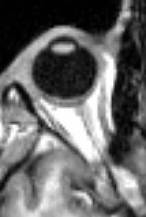
Oculomotor muscles
| Extraocular muscles | |
|---|---|

|
|
| Details | |
| System | Visual system |
| Origin | Annulus of Zinn, maxillary and sphenoid bone |
| Insertion | Tarsal plate of upper eyelid, eye |
| Artery |
Ophthalmic artery, lacrimal artery, infraorbital artery, anterior ciliary arteries, superior and inferior orbital veins |
| Nerve | Oculomotor, trochlear and Abducens nerve |
| Actions | See table |
| Identifiers | |
| Latin | Musculi externi bulbi oculi |
| MeSH | D009801 |
| TA | A04.1.01.001 |
| FMA | 49033 71101, 49033 |
|
Anatomical terms of muscle
[]
|
|
The extraocular muscles are the six muscles that control movement of the eye and one muscle that controls eyelid elevation (levator palpebrae). The actions of the six muscles responsible for eye movement depend on the position of the eye at the time of muscle contraction.
Since only a small part of the eye called the fovea provides sharp vision, the eye must move to follow a target. Eye movements must be precise and fast. This is seen in scenarios like reading, where the reader must shift gaze constantly. Although under voluntary control, most eye movement is accomplished without conscious effort. Precisely how the integration between voluntary and involuntary control of the eye occurs is a subject of continuing research. It is known, however, that the vestibulo-ocular reflex plays an important role in the involuntary movement of the eye.
Four of the extraocular muscles have their origin in the back of the orbit in a fibrous ring called the annulus of Zinn: the four rectus muscles. The four rectus muscles attach directly to the front half of the eye (anterior to the eye's equator), and are named after their straight paths. Note that medial and lateral are relative terms. Medial indicates near the midline, and lateral describes a position away from the midline. Thus, the medial rectus is the muscle closest to the nose. The superior and inferior recti do not pull straight back on the eye, because both muscles also pull slightly medially. This posterior medial angle causes the eye to roll with contraction of either the superior rectus or inferior rectus muscles. The extent of rolling in the recti is less than the oblique, and opposite from it.
The superior oblique muscle originates at the back of the orbit (a little closer to the medial rectus, though medial to it, getting rounder as it courses forward to a rigid, cartilaginous pulley, called the trochlea, on the upper, nasal wall of the orbit. The muscle becomes tendinous about 10mm before it passes through the pulley, turning sharply across the orbit, and inserts on the lateral, posterior part of the globe. Thus, the superior oblique travels posteriorly for the last part of its path, going over the top of the eye. Due to its unique path, the superior oblique, when activated, pulls the eye downward and laterally.
...
Wikipedia
