Ophthalmic artery
| Ophthalmic artery | |
|---|---|

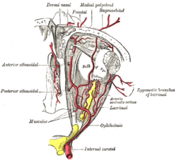
The ophthalmic artery and its branches.
|
|

Circle of Willis (Ophthalmic artery labeled at upper right)
|
|
| Details | |
| Source | Internal carotid |
| Branches |
Lacrimal artery Supraorbital artery Posterior ethmoidal artery Anterior ethmoidal artery Internal palpebral artery Supratrochlear artery Dorsal nasal artery Long posterior ciliary arteries Short posterior ciliary arteries Anterior ciliary artery Central retinal artery Superior muscular artery Inferior muscular artery |
| Vein | superior ophthalmic, inferior ophthalmic |
| Identifiers | |
| Latin | arteria ophthalmica |
| MeSH | A07.231.114.622 |
| TA | A12.2.06.016 |
| FMA | 49868 |
|
Anatomical terminology []
|
|
The ophthalmic artery (OA) is the first branch of the internal carotid artery distal to the cavernous sinus. Branches of the OA supply all the structures in the orbit as well as some structures in the nose, face and meninges. Occlusion of the OA or its branches can produce sight-threatening conditions.
The OA emerges from the internal carotid artery usually just after the latter emerges from the cavernous sinus although in some cases, the OA branches just before the internal carotid exits the cavernous sinus. The OA arises from the internal carotid along the medial side of the anterior clinoid process and runs anteriorly passing through the optic canal with and inferolaterally to the optic nerve. Here, it should be noted that the ophthalmic artery can also pass superiorly to the optic nerve in a minority of cases. In the posterior third of the cone of the orbit, the ophthalmic artery turns sharply medially to run along the medial wall of the orbit.
The central retinal artery is the first, and one of the smaller branches of the OA and runs in the dura mater inferior to the optic nerve. About 12.5mm (0.5 inch) posterior to the globe, the central retinal artery turns superiorly and penetrates the optic nerve continuing along the center of the optic nerve entering the eye to supply the inner retinal layers.
The next branch of the OA is the lacrimal artery, one of the largest, arises just as the OA enters the orbit and runs along the superior edge of the lateral rectus muscle to supply the lacrimal gland, eyelids and conjunctiva.
The OA then turns medially giving off 1 to 5 posterior ciliary arteries (PCA) that subsequently branch into the long and short posterior ciliary arteries (LPCA and SPCA respectively) which perforate the sclera posteriorly in the vicinity of the optic nerve and macula to supply the posterior uveal tract. In the past, anatomists made little distinction between the posterior ciliary arteries and the short and long posterior ciliary arteries often using the terms synonymously. However, recent work by Hayreh has shown that there is both an anatomic and clinically useful distinction. The PCAs arise directly from the OA and are end arteries which is to say no PCA or any of its branches anastomose with any other artery. Consequently, sudden occlusion of any PCA will produce an infarct in the region of the choroid supplied by that particular PCA. Occlusion of a short or long PCA will produce a smaller choroidal infarct within the larger area supplied by the specific parent PCA.
...
Wikipedia
