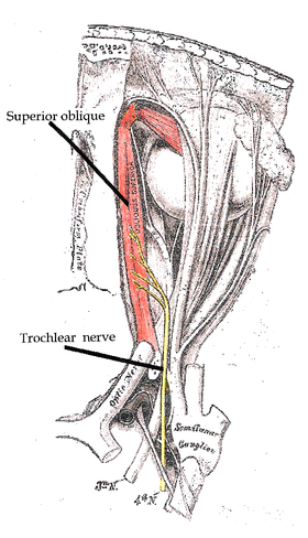
Trochlear nerve
| Trochlear nerve | |
|---|---|

Path of the Trochlear nerve, right eye, superior view
|
|

Inferior view of the human brain, with the cranial nerves labelled.
|
|
| Details | |
| Innervates | superior oblique muscle |
| Identifiers | |
| Latin | nervus trochlearis |
| MeSH | A08.800.800.120.800 |
| TA | A14.2.01.011 |
| FMA | 50865 |
|
Anatomical terms of neuroanatomy
[]
|
|
The trochlear nerve, also called the fourth cranial nerve or cranial nerve IV, is a motor nerve (a somatic efferent nerve) that innervates only a single muscle: the superior oblique muscle of the eye, which operates through the pulley-like trochlea.
The trochlear nerve is unique among the cranial nerves in several respects:
Homologous trochlear nerves are found in all jawed vertebrates. The unique features of the trochlear nerve, including its dorsal exit from the brainstem and its contralateral innervation, are seen in the primitive brains of sharks.
The human trochlear nerve is derived from the basal plate of the embryonic midbrain.
The trochlear nerve emerges from the dorsal aspect of the brainstem at the level of the caudal mesencephalon, just below the inferior colliculus. It circles anteriorly around the brainstem and runs forward toward the eye in the subarachnoid space. It passes between the posterior cerebral artery and the superior cerebellar artery, and then pierces the dura just under free margin of the tentorium cerebelli, close to the crossing of the attached margin of the tentorium and within millimeters of the posterior clinoid process. It runs on the lateral wall of the cavernous sinus, where it is joined by the other two extraocular nerves (oculomotor - cranial nerve III and abducens - cranial nerve VI) and the first two branches of the trigeminal nerve (V), Ophthalmic (V1) and Maxillary (V2). The internal carotid artery also runs within the cavernous sinus. Finally, it enters the orbit through the superior orbital fissure and innervates the superior oblique muscle.
...
Wikipedia
