Maxillary bone
| Maxilla | |
|---|---|

Side view. Maxilla visible at bottom left, in green.
|
|

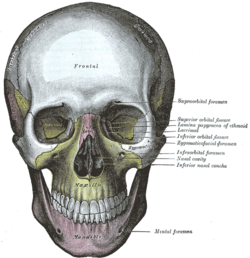
Front view. Maxilla visible at center, in yellow.
|
|
| Details | |
| Precursor | 1st branchial arch |
| Identifiers | |
| MeSH | A02.835.232.781.324.502.645 |
| FMA | 9711 |
|
Anatomical terms of bone
[]
|
|
The maxilla (plural: maxillae /mækˈsɪliː/) is the upper jawbone formed from the fusion of two maxillary bones. The upper jaw includes the palate of the mouth. The two maxillary bones are fused at the inter maxillary suture. This is similar to the mandible (lower jaw), which is also a fusion of two mandibular bones at the mandibular symphysis.
Sometimes as in bony fish, the maxilla is called the "upper maxilla", with the mandible being called the "lower maxilla". Conversely, in birds the upper jaw is often called the "upper mandible".
Each maxillary bone consists of:
Each maxilla articulates with nine bones:
Sometimes it articulates with the orbital surface, and sometimes with the lateral pterygoid plate of the sphenoid.
The maxilla is ossified in membrane. Mall and Fawcett maintain that it is ossified from two centers only, one for the maxilla proper and one for the premaxilla.
These centers appear during the sixth week of prenatal development and unite in the beginning of the third month, but the suture between the two portions persists on the palate until nearly middle life. Mall states that the frontal process is developed from both centers.
The maxillary sinus appears as a shallow groove on the nasal surface of the bone about the fourth month of development, but does not reach its full size until after the second dentition.
The maxilla was formerly described as ossifying from six centers, viz.,
At birth the transverse and antero-posterior diameters of the bone are each greater than the vertical.
The frontal process is well-marked and the body of the bone consists of little more than the alveolar process, the teeth sockets reaching almost to the floor of the orbit.
...
Wikipedia
