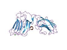
Laminins are high-molecular weight (~400 to ~900 kDa) proteins of the extracellular matrix. They are a major component of the basal lamina (one of the layers of the basement membrane), a protein network foundation for most cells and organs. The laminins are an important and biologically active part of the basal lamina, influencing cell differentiation, migration, and adhesion.
Laminins are heterotrimeric proteins that contain an α-chain, a β-chain, and a γ-chain, found in five, four, and three genetic variants, respectively. The laminin molecules are named according to their chain composition. Thus, laminin-511 contains α5, β1, and γ1 chains. Fourteen other chain combinations have been identified in vivo. The trimeric proteins intersect to form a cross-like structure that can bind to other cell membrane and extracellular matrix molecules. The three shorter arms are particularly good at binding to other laminin molecules, which allows them to form sheets. The long arm is capable of binding to cells, which helps anchor organized tissue cells to the membrane.
The laminin family of glycoproteins are an integral part of the structural scaffolding in almost every tissue of an organism. They are secreted and incorporated into cell-associated extracellular matrices. Laminin is vital for the maintenance and survival of tissues. Defective laminins can cause muscles to form improperly, leading to a form of muscular dystrophy, lethal skin blistering disease (junctional epidermolysis bullosa) and defects of the kidney filter (nephrotic syndrome).
Fifteen laminin trimers have been identified. The laminins are combinations of different alpha-, beta-, and gamma-chains.
Laminins were previously numbered - e.g. laminin-1, laminin-2, laminin-3 - but the nomenclature was recently changed to describe which chains are present in each isoform.
Laminins form independent networks and are associated with type IV collagen networks via entactin,fibronectin, and perlecan. They also bind to cell membranes through integrin receptors and other plasma membrane molecules, such as the dystroglycan glycoprotein complex and Lutheran blood group glycoprotein. Through these interactions, laminins critically contribute to cell attachment and differentiation, cell shape and movement, maintenance of tissue phenotype, and promotion of tissue survival. Some of these biological functions of laminin have been associated with specific amino-acid sequences or fragments of laminin. For example, the peptide sequence [GTFALRGDNGDNGQ], which is located on the alpha-chain of laminin, promotes adhesion of endothelial cells.
...
Wikipedia


