CD25+
| interleukin 2 receptor, alpha chain | |
|---|---|
 |
|
| Identifiers | |
| Symbol | IL2RA |
| Alt. symbols | IL2R CD25 |
| Entrez | 3559 |
| HUGO | 6008 |
| OMIM | 147730 |
| RefSeq | NM_000417 |
| UniProt | P01589 |
| Other data | |
| Locus | Chr. 10 p15.1 |
| interleukin 2 receptor, beta chain | |
|---|---|
 |
|
| Identifiers | |
| Symbol | IL2RB |
| Alt. symbols | CD122 |
| Entrez | 3560 |
| HUGO | 6009 |
| OMIM | 146710 |
| RefSeq | NM_000878 |
| UniProt | P14784 |
| Other data | |
| Locus | Chr. 22 q13 |
| interleukin 2 receptor, gamma chain (severe combined immunodeficiency) | |
|---|---|
 |
|
| Identifiers | |
| Symbol | IL2RG |
| Alt. symbols | SCIDX1, IMD4, CD132 |
| Entrez | 3561 |
| HUGO | 6010 |
| OMIM | 308380 |
| RefSeq | NM_000206 |
| UniProt | P31785 |
| Other data | |
| Locus | Chr. X q13 |
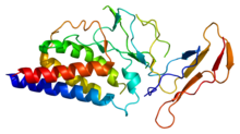
The interleukin-2 receptor (IL-2R) is a heterotrimeric protein expressed on the surface of certain immune cells, such as lymphocytes, that binds and responds to a cytokine called IL-2.
IL-2 binds to the IL-2 receptor, which has three forms, generated by different combinations of three different proteins, often referred to as "chains": α (alpha) (also called IL-2Rα, CD25, or Tac antigen), β (beta) (also called IL-2Rβ, or CD122), and γ (gamma) (also called IL-2Rγ, γc, common gamma chain, or CD132); these subunits are also parts of receptors for other cytokines. The β and γ chains of the IL-2R are members of the type I cytokine receptor family.
The three receptor chains are expressed separately and differently on various cell types and can assemble in different combinations and orders to generate low, intermediate, and high affinity IL-2 receptors.
The α chain binds IL-2 with low affinity, the combination of β and γ together form a complex that binds IL-2 with intermediate affinity, primarily on memory T cells and NK cells; and all three receptor chains form a complex that binds IL-2 with high affinity (Kd ~ 10−11 M) on activated T cells and regulatory T cells The intermediate and high affinity receptor forms are functional and cause changes in the cell when IL-2 binds to them.
The structure of the stable complex formed when IL-2 binds to the high affinity receptor has been determined using X-ray crystallography. The structure supports a model wherein IL-2 initially binds to the α chain, then the β is recruited, and finally γ.
The three IL-2 receptor chains span the cell membrane and extend into the cell, thereby delivering biochemical signals to the cell interior. The alpha chain does not participate in signaling, but the beta chain is complexed with an enzyme called Janus kinase 1 (JAK1), that is capable of adding phosphate groups to molecules. Similarly the gamma chain complexes with another tyrosine kinase called JAK3. These enzymes are activated by IL-2 binding to the external domains of the IL-2R. As a consequence, three intracellular signaling pathways are initiated, the MAP kinase pathway, the Phosphoinositide 3-kinase (PI3K) pathway, and the JAK-STAT pathway.
...
Wikipedia
