Arginase
| Arginase | |||||||||
|---|---|---|---|---|---|---|---|---|---|

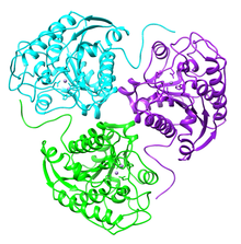
Ribbon diagram of human arginase I trimer. PDB entry 2pha
|
|||||||||
| Identifiers | |||||||||
| EC number | 3.5.3.1 | ||||||||
| CAS number | 9000-96-8 | ||||||||
| Databases | |||||||||
| IntEnz | IntEnz view | ||||||||
| BRENDA | BRENDA entry | ||||||||
| ExPASy | NiceZyme view | ||||||||
| KEGG | KEGG entry | ||||||||
| MetaCyc | metabolic pathway | ||||||||
| PRIAM | profile | ||||||||
| PDB structures | RCSB PDB PDBe PDBsum | ||||||||
| Gene Ontology | AmiGO / EGO | ||||||||
|
|||||||||
| Search | |
|---|---|
| PMC | articles |
| PubMed | articles |
| NCBI | proteins |
| Liver arginase | |
|---|---|
 |
|
| Identifiers | |
| Symbol | ARG1 |
| Entrez | 383 |
| HUGO | 663 |
| OMIM | 608313 |
| RefSeq | NM_000045 |
| UniProt | P05089 |
| Other data | |
| EC number | 3.5.3.1 |
| Locus | Chr. 6 q23 |
| Arginase, type II | |
|---|---|
| Identifiers | |
| Symbol | ARG2 |
| Entrez | 384 |
| HUGO | 664 |
| OMIM | 107830 |
| RefSeq | NM_001172 |
| UniProt | P78540 |
| Other data | |
| EC number | 3.5.3.1 |
| Locus | Chr. 14 q24.1 |
Arginase (EC 3.5.3.1, arginine amidinase, canavanase, L-arginase, arginine transamidinase) is a manganese-containing enzyme. The reaction catalyzed by this enzyme is: arginine + H2O → ornithine + urea. It is the final enzyme of the urea cycle. It is ubiquitous to all domains of life.
Arginase belong to the ureohydrolase family of enzymes.
Arginase catalyzes the fifth and final step in the urea cycle, a series of biochemical reactions in mammals during which the body disposes of harmful ammonia. Specifically, arginase converts L-arginine into L-ornithine and urea. Mammalian arginase is active as a trimer, but some bacterial arginases are hexameric. The enzyme requires a two-molecule metal cluster of manganese in order to maintain proper function. These Mn2+ions coordinate with water, orienting and stabilizing the molecule and allowing water to act as a nucleophile and attack L-arginine, hydrolyzing it into ornithine and urea.
In most mammals, two isozymes of this enzyme exist; the first, Arginase I, functions in the urea cycle, and is located primarily in the cytoplasm of the liver. The second isozyme, Arginase II, has been implicated in the regulation of the arginine/ornithine concentrations in the cell. It is located in mitochondria of several tissues in the body, with most abundance in the kidney and prostate. It may be found at lower levels in macrophages, lactating mammary glands, and brain. The second isozyme may be found in the absence of other urea cycle enzymes.
The active site holds L-arginine in place via hydrogen bonding between the guanidine chloride group with Glu227. This bonding orients L-arginine for nucleophilic attack by the metal-associated hydroxide ion at the guanidine chloride group. This results in a tetrahedral intermediate. The manganese ions act to stabilize both the hydroxyl group in the tetrahedral intermediate, as well as the developing sp3 lone electron pair on the NH2 group as the tetrahedral intermediate is formed.
...
Wikipedia
