Thalami
| Thalamus | |
|---|---|

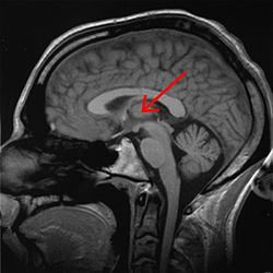
thalamus marked (MRI cross-section)
|
|

anterolateral view
|
|
| Details | |
| Part of | Diencephalon |
| Components | See List of thalamic nuclei |
| Artery | Posterior cerebral artery and branches |
| Identifiers | |
| Latin | thalamus dorsalis |
| MeSH | A08.186.211.730.385.826 |
| NeuroNames | hier-283 |
| NeuroLex ID | Thalamus |
| TA |
A14.1.08.101 A14.1.08.601 |
| FMA | 62007 |
|
Anatomical terms of neuroanatomy
[]
|
|
The thalamus (from Greek , "chamber") is the large mass of gray matter in the dorsal part of the diencephalon of the brain with several functions such as relaying of sensory and motor signals to the cerebral cortex, and the regulation of consciousness, sleep, and alertness.
It is a symmetrical structure of two halves, within the vertebrate brain, situated between the cerebral cortex and the midbrain. The medial surface of the two halves constitute the upper lateral wall of the third ventricle.
It is the main product of the embryonic diencephalon, as first assigned by Wilhelm His, Sr. in 1893.
The thalamus is located in the forebrain which is superior to the midbrain, near the center of the brain, with nerve fibers projecting out to the cerebral cortex in all directions. The medial surface of the thalamus constitutes the upper part of the lateral wall of the third ventricle, and is connected to the corresponding surface of the opposite thalamus by a flattened gray band, the interthalamic adhesion.
The thalamus derives its blood supply from a number of arteries: the polar artery (posterior communicating artery), paramedian thalamic-subthalamic arteries, inferolateral (thalamogeniculate) arteries, and posterior (medial and lateral) choroidal arteries. These are all branches of the posterior cerebral artery.
Some people have the artery of Percheron, which is a rare anatomic variation in which a single arterial trunk arises from the posterior cerebral artery to supply both parts of the thalamus.
...
Wikipedia
