Gray matter
| Grey matter | |
|---|---|

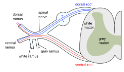
The formation of the spinal nerve from the dorsal and ventral roots. (Grey matter labeled at center right.)
|
|

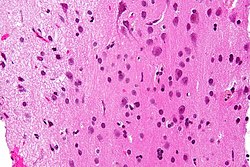
Micrograph showing grey matter, with the characteristic neuronal cell bodies (dark shade of pink), and white matter with its characteristic fine meshwork-like appearance (left of image - lighter shade of pink). HPS stain.
|
|
| Details | |
| Identifiers | |
| Latin | Substantia grisea |
| Dorlands /Elsevier |
Grey matter |
| TA | A14.1.00.002 |
| FMA | 67242 |
|
Anatomical terminology
[]
|
|
Grey matter (or gray matter) is a major component of the central nervous system, consisting of neuronal cell bodies, neuropil (dendrites and myelinated as well as unmyelinated axons), glial cells (astroglia and oligodendrocytes), synapses, and capillaries. Grey matter is distinguished from white matter, in that it contains numerous cell bodies and relatively few myelinated axons, while white matter contains relatively very few cell bodies and is composed chiefly of long-range myelinated axon tracts. The colour difference arises mainly from the whiteness of myelin. In living tissue, grey matter actually has a very light grey colour with yellowish or pinkish hues, which come from capillary blood vessels and neuronal cell bodies.
Grey matter refers to unmyelinated neurons and other cells of the central nervous system. It is present in the brain, brainstem and cerebellum, and present throughout the spinal cord.
Grey matter is distributed at the surface of the cerebral hemispheres (cerebral cortex) and of the cerebellum (cerebellar cortex), as well as in the depths of the cerebrum (thalamus; hypothalamus; subthalamus, basal ganglia – putamen, globus pallidus, nucleus accumbens; septal nuclei), cerebellar (deep cerebellar nuclei – dentate nucleus, globose nucleus, emboliform nucleus, fastigial nucleus), brainstem (substantia nigra, red nucleus, olivary nuclei, cranial nerve nuclei).
...
Wikipedia
