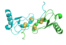
Macrophage inflammatory protein
| chemokine (C-C motif) ligand 3 | |
|---|---|

|
|
| Identifiers | |
| Symbol | CCL3 |
| Alt. symbols | SCYA3, MIP-1α |
| Entrez | 6348 |
| HUGO | 10627 |
| OMIM | 182283 |
| PDB | 1B50 More structures |
| RefSeq | NM_002983 |
| UniProt | P10147 |
| Other data | |
| Locus | Chr. 17 q12 |
| chemokine (C-C motif) ligand 4 | |
|---|---|

|
|
| Identifiers | |
| Symbol | CCL4 |
| Alt. symbols | SCYA4, MIP-1β, LAG1 |
| Entrez | 6351 |
| HUGO | 10630 |
| OMIM | 182284 |
| PDB | 1HUM More structures |
| RefSeq | NM_002984 |
| UniProt | P13236 |
| Other data | |
| Locus | Chr. 17 q21-q23 |
Macrophage Inflammatory Proteins (MIP) belong to the family of chemotactic cytokines known as chemokines. In humans, there are two major forms, MIP-1α and MIP-1β that are now officially named CCL3 and CCL4, respectively. Both are major factors produced by macrophages after they are stimulated with bacterial endotoxins. They are crucial for immune responses towards infection and inflammation. They activate human granulocytes (neutrophils, eosinophils and basophils) which can lead to acute neutrophilic inflammation. They also induce the synthesis and release of other pro-inflammatory cytokines such as interleukin 1 (IL-1), IL-6 and TNF-α from fibroblasts and macrophages. The genes for CCL3 and CCL4 are both located on human chromosome 17.
They are produced by many cells, particularly macrophages, dendritic cells, and lymphocytes. MIP-1 are best known for their chemotactic and proinflammatory effects but can also promote homoeostasis. Biophysical analyses and mathematical modelling has shown that MIP-1 reversibly forms a polydisperse distribution of rod-shaped polymers in solution. Polymerization buries receptor-binding sites of MIP-1, thus depolymerization mutations enhance MIP-1 to arrest monocytes onto activated human endothelium.
...
Wikipedia
