FERM domain
In molecular biology, the FERM domain (F for 4.1 protein, E for ezrin, R for radixin and M for moesin) is a widespread protein module involved in localising proteins to the plasma membrane. FERM domains are found in a number of cytoskeletal-associated proteins that associate with various proteins at the interface between the plasma membrane and the cytoskeleton. The FERM domain is located at the N terminus in the majority of proteins in which it is found.
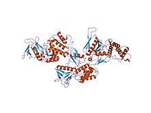
Ezrin, moesin, and radixin are highly related proteins (ERM protein family), but the other proteins in which the FERM domain is found do not share any region of similarity outside of this domain. ERM proteins are made of three domains, the FERM domain, a central helical domain and a C-terminal tail domain, which binds F-actin. The amino-acid sequence of the FERM domain is highly conserved among ERM proteins and is responsible for membrane association by direct binding to the cytoplasmic domain or tail of integral membrane proteins. ERM proteins are regulated by an intramolecular association of the FERM and C-terminal tail domains that masks their binding sites for other molecules. For cytoskeleton-membrane cross-linking, the dormant molecules becomes activated and the FERM domain attaches to the membrane by binding specific membrane proteins, while the last 34 residues of the tail bind actin filaments. Aside from binding to membranes, the activated FERM domain of ERM proteins can also bind the guanine nucleotide dissociation inhibitor of Rho GTPase (RhoDGI), which suggests that in addition to functioning as a cross-linker, ERM proteins may influence Rho signalling pathways. The crystal structure of the FERM domain reveals that it is composed of three structural modules (F1, F2, and F3) that together form a compact clover-shaped structure. The N-terminal module is ubiquitin-like. The C-terminal module is a PH-like domain.
...
Wikipedia


