Corticoreticular fibers
| Reticular formation | |
|---|---|

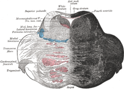
Axial section of the pons, at its upper part. (Formatio reticularis labeled at left.)
|
|

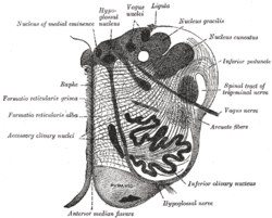
Section of the medulla oblongata at about the middle of the olive. (Formatio reticularis grisea and formatio reticularis alba labeled at left.)
|
|
| Details | |
| Identifiers | |
| Latin | formatio reticularis |
| MeSH | A08.186.211.132.772 |
| NeuroNames | ancil-225 |
| NeuroLex ID | Reticular formation |
| Dorlands /Elsevier |
f_13/12374790 |
| TA |
A14.1.00.021 A14.1.05.403 A14.1.06.327 |
| FMA | 77719 |
|
Anatomical terms of neuroanatomy
[]
|
|
The reticular formation is a set of interconnected nuclei that are located throughout the brainstem. The reticular formation is not anatomically well defined because it includes neurons located in diverse parts of the brain. The neurons of the reticular formation all play a crucial role in maintaining behavioral arousal and consciousness. The functions of the reticular formation are modulatory and premotor. The modulatory functions are primarily found in the rostral sector of the reticular formation and the premotor functions are localized in the neurons in more caudal regions.
The reticular formation is divided into three columns: raphe nuclei (median), (medial zone), and parvocellular reticular nuclei (lateral zone). The raphe nuclei are the place of synthesis of the neurotransmitter serotonin, which plays an important role in mood regulation. The gigantocellular nuclei are involved in motor coordination. The parvocellular nuclei regulate exhalation.
It is essential for governing some of the basic functions of higher organisms and is one of the phylogenetically oldest portions of the brain.
The reticular formation has been functionally cleaved both sagittally and coronally.
Traditionally the nuclei are divided into three columns
The original functional differentiation was a division of caudal and rostral, this was based upon the observation that the lesioning of the rostral reticular formation induces a hypersomnia in the cat brain. In contrast, lesioning of the more caudal portion of the reticular formation produces insomnia in cats. This study has led to the idea that the caudal portion inhibits the rostral portion of the reticular formation.
Sagittal division reveals more morphological distinctions. The raphe nuclei form a ridge in the middle of the reticular formation, and, directly to its periphery, there is a division called the medial reticular formation. The medial RF is large and has long ascending and descending fibers, and is surrounded by the lateral reticular formation. The lateral RF is close to the motor nuclei of the cranial nerves, and mostly mediates their function.
...
Wikipedia
