Raphe nuclei
| Raphe nuclei | |
|---|---|

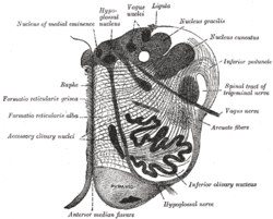
Section of the medulla oblongata at about the middle of the olive. (Raphe nuclei not labeled, but 'raphe' labeled at left.)
|
|

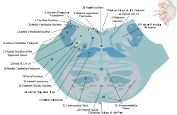
Horizontal cross section of the brainstem at the lower pons. The raphe nucleus is labeled #18 in the middle.
|
|
| Details | |
| Identifiers | |
| Latin | nuclei raphe |
| MeSH | A08.186.211.132.659.632 |
| NeuroLex ID | Raphe Nuclei |
| TA |
A14.1.04.257 A14.1.04.318 A14.1.05.402 A14.1.05.601 A14.1.06.401 |
| FMA | 84017 |
|
Anatomical terms of neuroanatomy
[]
|
|
The raphe nuclei (Greek: ῥαφή "seam") are a moderate-size cluster of nuclei found in the brain stem. Their main function is to release serotonin to the rest of the brain.Selective serotonin reuptake inhibitor (SSRI) antidepressants are believed to act in these nuclei, as well as at their targets.
The raphe nuclei are traditionally considered to be the medial portion of the reticular formation, and appear as a ridge of cells in the center and most medial portion of the brain stem.
In order from caudal to rostral, the raphe nuclei are known as the nucleus raphe obscurus, the nucleus raphe pallidus, the nucleus raphe magnus, the nucleus raphe pontis, the median raphe nucleus, dorsal raphe nucleus, caudal linear nucleus. In the first systematic examination of the raphe nuclei, Taber et al.. (1960) originally proposed the existence of two linear nuclei (nucleus linearis intermedius and nucleus linearis rostralis). This study was published before techniques enabling the visualization of serotonin or the enzymes participating in its synthesis had been developed, as first demonstrated by Dahlström and Fuxe in 1964. Later, it was revealed that of these two nuclei, only the former (nucleus linearis intermedius, now known as the caudal linear nucleus), proved to contain serotonin-producing neurons, though both of them contain dopaminergic neurons.
...
Wikipedia
