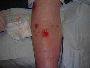
Venous ulcer
| Venous ulcer | |
|---|---|
 |
|
| Venous ulcer on the back of the right leg. | |
| Classification and external resources | |
| Specialty | dermatology |
| ICD-10 | I83.0, I83.2, L97 |
| ICD-9-CM | 454.0 |
| DiseasesDB | 29114 |
| MedlinePlus | 000834 |
| MeSH | D014647 |
Venous ulcers (venous insufficiency ulceration, stasis ulcers, stasis dermatitis, varicose ulcers, or ulcus cruris) are wounds that are thought to occur due to improper functioning of venous valves, usually of the legs (hence leg ulcers). They are the major occurrence of chronic wounds, occurring in 70% to 90% of leg ulcer cases. Venous ulcers develop mostly along the medial distal leg, and can be very painful.
Edema and fibrinous exudate leads to fibrosis of subcutaneous tissues with localized pigment loss and dilation of capillary loops. This is called atrophic blanche. This can occur around ankles and gives an appearance of inverted champagne bottle to legs. Large ulcers may encircle the leg. Lymphedema results from obliteration of superficial lymphatics. There is hypertrophy of overlying epidermis giving polypoid appearance, known as lipodermatosclerosis.
Venous ulcer before surgery
Healing process of a chronic venous stasis ulcer of the lower leg
Healing venous ulcer after one month
The exact cause of venous ulcers is not certain, but they are thought to arise when venous valves that exist to prevent backflow of blood do not function properly, causing the pressure in veins to increase. The body needs the pressure gradient between arteries and veins in order for the heart to pump blood forward through arteries and into veins. When venous hypertension exists, arteries no longer have significantly higher pressure than veins, and blood is not pumped as effectively into or out of the area.
Venous hypertension may also stretch veins and allow blood proteins to leak into the extravascular space, isolating extracellular matrix (ECM) molecules and growth factors, preventing them from helping to heal the wound. Leakage of fibrinogen from veins as well as deficiencies in fibrinolysis may also cause fibrin to build up around the vessels, preventing oxygen and nutrients from reaching cells. Venous insufficiency may also cause white blood cells (leukocytes) to accumulate in small blood vessels, releasing inflammatory factors and reactive oxygen species (ROS, free radicals) and further contributing to chronic wound formation. Buildup of white blood cells in small blood vessels may also plug the vessels, further contributing to ischemia. This blockage of blood vessels by leukocytes may be responsible for the "no reflow phenomenon," in which ischemic tissue is never fully reperfused. Allowing blood to flow back into the limb, for example by elevating it, is necessary but also contributes to reperfusion injury. Other comorbidities may also be the root cause of venous ulcers.
...
Wikipedia
