UDP-glucose pyrophosphorylase
| UTP—glucose-1-phosphate uridylyltransferase | |||||||||
|---|---|---|---|---|---|---|---|---|---|

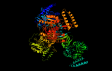
Human UTP—glucose-1-phosphate uridylyltransferase cartoon created in pymol
|
|||||||||
| Identifiers | |||||||||
| EC number | 2.7.7.9 | ||||||||
| CAS number | 9026-22-6 | ||||||||
| Databases | |||||||||
| IntEnz | IntEnz view | ||||||||
| BRENDA | BRENDA entry | ||||||||
| ExPASy | NiceZyme view | ||||||||
| KEGG | KEGG entry | ||||||||
| MetaCyc | metabolic pathway | ||||||||
| PRIAM | profile | ||||||||
| PDB structures | RCSB PDB PDBe PDBsum | ||||||||
| Gene Ontology | AmiGO / EGO | ||||||||
|
|||||||||
| Search | |
|---|---|
| PMC | articles |
| PubMed | articles |
| NCBI | proteins |
| UDP–glucose pyrophosphorylase 1 | |
|---|---|
| Identifiers | |
| Symbol | UGP1 |
| Entrez | 7359 |
| HUGO | 12526 |
| OMIM | 191750 |
| Other data | |
| EC number | 2.7.7.9 |
| Locus | Chr. 1 q21-q22 |
| UDP–glucose pyrophosphorylase 2 | |
|---|---|
| Identifiers | |
| Symbol | UGP2 |
| Entrez | 7360 |
| HUGO | 12527 |
| OMIM | 191760 |
| RefSeq | NM_006759 |
| UniProt | Q16851 |
| Other data | |
| EC number | 2.7.7.9 |
| Locus | Chr. 2 p14-p13 |
UTP—glucose-1-phosphate uridylyltransferase also known as glucose-1-phosphate uridylyltransferase (or UDP–glucose pyrophosphorylase) is an enzyme involved in carbohydrate metabolism. It synthesizes UDP-glucose from glucose-1-phosphate and UTP; i.e.,
UTP—glucose-1-phosphate uridylyltransferase is an enzyme found in all three domains (bacteria, eukarya, and archaea) as it is a key player in glycogenesis and cell wall synthesis. Its role in sugar metabolism has been studied extensively in plants in order to understand plant growth and increase agricultural production. Recently, human UTP—glucose-1-phosphate uridylyltransferase has been studied and crystallized, revealing a different type of regulation than other organisms previously studied. Its significance is derived from the many uses of UDP-glucose including galactose metabolism, glycogen synthesis, glycoprotein synthesis, and glycolipid synthesis.
The structure of UTP—glucose-1-phosphate uridylyltransferase is significantly different between prokaryotes and eukaryotes, but within eukaryotes, the primary, secondary, and tertiary structures of the enzyme are quite conserved. In many species, UTP—glucose-1-phosphate uridylyltransferase is found as a homopolymer consisting of identical subunits in a symmetrical quaternary structure. The number of subunits varies across species: for instance, in Escherichia coli, the enzyme is found as a tetramer, whereas in Burkholderia xenovorans, the enzyme is dimeric. In humans and in yeast, the enzyme is active as an octamer consisting of two tetramers stacked onto one another with conserved hydrophobic residues at the interfaces between the subunits. In contrast, the enzyme in plants has conserved charged residues forming the interface between subunits.
...
Wikipedia
