Precuneus
| Precuneus | |
|---|---|

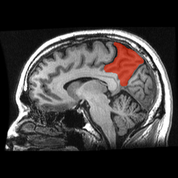
Medial surface of left cerebral hemisphere. (Precuneus visible at top left.)
|
|

Sagittal MRI slice with the precuneus shown in red.
|
|
| Details | |
| Identifiers | |
| Latin | Praecuneus |
| NeuroNames | hier-92 |
| NeuroLex ID | Precuneus cortex |
| TA | A14.1.09.223 |
| FMA | 61900 |
|
Anatomical terms of neuroanatomy
[]
|
|
The precuneus is a part of the superior parietal lobule forward of the occipital lobe (cuneus). It is hidden in the medial longitudinal fissure between the two cerebral hemispheres. It is sometimes described as the medial area of the superior parietal cortex. The precuneus is bounded anteriorly by the marginal branch of the cingulate sulcus, posteriorly by the parietooccipital sulcus, and inferiorly by the subparietal sulcus. It is involved with episodic memory, visuospatial processing, reflections upon self, and aspects of consciousness.
The location of the precuneus makes it difficult to study. Furthermore, it is rarely subject to isolated injury due to strokes, or trauma such as gunshot wounds. This has resulted in it being "one of the less accurately mapped areas of the whole cortical surface". While originally described as homogeneous by Korbinian Brodmann, it is now appreciated to contain three subdivisions.
It is also known after Achille-Louis Foville as the quadrate lobule of Foville. The Latin form of praecuneus was first used in 1868 and the English precuneus in 1879.
The precuneus is located on the inside between the two cerebral hemispheres in the rear region between the somatosensory cortex and forward of the cuneus (which contains the visual cortex). It is above the posterior cingulate. Following Korbinian Brodmann it has traditionally been considered a homogeneous structure and with limited distinction between it and the neighboring posterior cingulate area. Brodmann mapped it as the medial continuation of lateral parietal area 7.
...
Wikipedia
