Parieto-occipital sulcus
| Parieto-occipital sulcus | |
|---|---|

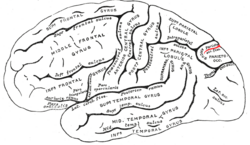
Fig. 726: Lateral surface of left cerebral hemisphere, viewed from the side.
|
|

Fig. 727: Medial surface of left cerebral hemisphere.
|
|
| Details | |
| Identifiers | |
| Latin | sulcus parietooccipitalis, fissura parietooccipitalis |
| NeuroNames | hier-33 |
| NeuroLex ID | Parieto-occipital sulcus |
| TA | A14.1.09.108 |
| FMA | 83754 |
|
Anatomical terms of neuroanatomy
[]
|
|
Only a small part of the parieto-occipital sulcus, or parietooccipital fissure is seen on the lateral surface of the hemisphere, its chief part being on the medial surface.
The lateral part of the parieto-occipital sulcus (Fig. 726) is situated about 5 centimeters (cm) in front of the occipital pole of the hemisphere, and measures about 1.25 cm. in length.
The medial part of the parieto-occipital sulcus (Fig. 727) runs downward and forward as a deep cleft on the medial surface of the hemisphere, and joins the calcarine fissure below and behind the posterior end of the corpus callosum. In most cases it contains a submerged gyrus. The parieto-occipital sulcus marks the boundary between the cuneus and precuneus, and also between the parietal and occipital lobes.
The parieto-occipiatal lobe has been found in various neuroimaging studies, including PET (positron-emission-tomography) studies, and SPECT (single-photon emission computed tomography) studies, to be involved along with the dorsolateral prefrontal cortex during planning.
Animation of left cerebral hemisphere. Parieto-occipital sulcus shown in red.
Medial surface of right hemisphere. Parieto-occipital sulcus labeled at top right as "*"
Medial surface of left hemisphere. Parieto-occipital sulcus visible at top left.
Human brain dissection video (1 min 52 sec). Demonstrating location of parieto-occipital sulcus of left cerebral hemisphere.
This article incorporates text in the public domain from the 20th edition of Gray's Anatomy (1918)
...
Wikipedia
