Periaqueductal gray
| Periaqueductal gray | |
|---|---|

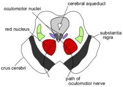
Section through superior colliculus showing path of oculomotor nerve. Periaqueductal gray is the gray area just peripheral to the cerebral aqueduct.
|
|

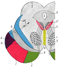
Transverse section through mid-brain.
|
|
| Details | |
| Identifiers | |
| Latin | Substantia grisea centralis |
| MeSH | A08.186.211.132.659.822.595 |
| NeuroNames | hier-501 |
| NeuroLex ID | Periaqueductal gray |
| TA | A14.1.06.321 |
| FMA | 83134 |
|
Anatomical terms of neuroanatomy
[]
|
|
The periaqueductal gray (PAG) (also known as the central gray) is the primary control center for descending pain modulation. It has enkephalin-producing cells that suppress pain.
The periaqueductal gray is the gray matter located around the cerebral aqueduct within the tegmentum of the midbrain. It projects to the nucleus raphe magnus, and also contains descending autonomic tracts. The ascending pain and temperature fibers of the spinothalamic tract send information to the PAG via the spinomesencephalic tract (so-named because the fibers originate in the spine and terminate in the PAG, in the mesencephalon or midbrain).
This region has been used as the target for brain-stimulating implants in patients with chronic pain.
Stimulation of the periaqueductal gray matter of the midbrain activates enkephalin-releasing neurons that project to the raphe nuclei in the brainstem. 5-HT (serotonin) released from the raphe nuclei descends to the dorsal horn of the spinal cord where it forms excitatory connections with the "inhibitory interneurons" located in Laminae II (aka the substantia gelatinosa). When activated, these interneurons release either enkephalin or dynorphin (endogenous opioid neurotransmitters), which bind to mu opioid receptors on the axons of incoming C and A-delta fibers carrying pain signals from nociceptors activated in the periphery. The activation of the mu-opioid receptor inhibits the release of substance P from these incoming first-order neurons and, in turn, inhibits the activation of the second-order neuron that is responsible for transmitting the pain signal up the spinothalamic tract to the ventroposteriolateral nucleus (VPL) of the thalamus. The nociceptive signal was inhibited before it was able to reach the cortical areas that interpret the signal as "pain" (such as the anterior cingulate). This is sometimes referred to as the Gate control theory of pain and is supported by the fact that electrical stimulation of the PAG results in immediate and profound analgesia. The periaqueductal gray is also activated by viewing distressing images associated with pain.
...
Wikipedia
