Mesothelia
| Mesothelium | |
|---|---|

|
|

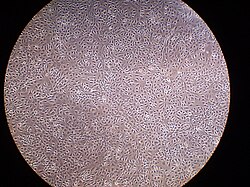
A layer of mesothelial cells grown in cell culture, featuring the typical "cobblestone" appearance
|
|
| Details | |
| Identifiers | |
| Latin | Mesothelium |
| MeSH | Mesothelium |
| TH | H2.00.02.0.02017, H3.04.08.0.00003 |
| FMA | 14074 |
|
Anatomical terminology
[]
|
|
The mesothelium is a membrane composed of simple squamous epithelium that forms the lining of several body cavities: the pleura (thoracic cavity), peritoneum (abdominal cavity including the mesentery), mediastinum and pericardium (heart sac). Mesothelial tissue also surrounds the male internal reproductive organs (the tunica vaginalis testis) and covers the internal reproductive organs of women (the tunica serosa uteri). Mesothelium that covers the internal organs is called visceral mesothelium, while the layer that covers the body walls is called the mesothelium. Mesothelium is the epithelial component of serosa.
Mesothelium derives from the embryonic mesoderm cell layer, that lines the coelom (body cavity) in the embryo. It develops into the layer of cells that covers and protects most of the internal organs of the body.
The mesothelium forms a monolayer of flattened squamous-like epithelial cells resting on a thin basement membrane supported by dense irregular connective tissue. Cuboidal mesothelial cells may be found at areas of injury, the milky spots of the omentum, and the peritoneal side of the diaphragm overlaying the lymphatic lacunae. The luminal surface is covered with microvilli. The proteins and serosal fluid trapped by the microvilli provide a slippery surface for internal organs to slide past one another.
...
Wikipedia
