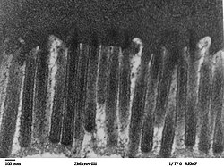
Microvilli
| Microvillus | |
|---|---|

Enterocytes with microvilli
|
|
| Details | |
| Identifiers | |
| Latin | Microvillus |
| Code | TH H1.00.01.1.01011 |
| TH | H1.00.01.1.01011 |
|
Anatomical terminology
[]
|
|
Microvilli (singular: microvillus) are microscopic cellular membrane protrusions that increase the surface area for diffusion and minimize any increase in volume, and are involved in a wide variety of functions, including absorption, secretion, cellular adhesion, and mechanotransduction.
Thousands of microvilli form a structure called the brush border that is found on the apical surface of some epithelial cells, such as the small intestines. (Microvilli should not be confused with intestinal villi, which are made of many cells. Each of these cells has many microvilli.) Microvilli are observed on the plasma surface of eggs, aiding in the anchoring of sperm cells that have penetrated the extracellular coat of egg cells. Clustering of elongated microtubules around a sperm allows for it to be drawn closer and held firmly so fusion can occur. They are large objects that increase surface area for absorption.
Microvilli are also of importance on the cell surface of white blood cells, as they aid in the migration of white blood cells.
Microvilli are covered in plasma membrane, which encloses cytoplasm and microfilaments. Though these are cellular extensions, there are little or no cellular organelles present in the microvilli.
Each microvillus has a dense bundle of cross-linked actin filaments, which serves as its structural core. 20 to 30 tightly bundled actin filaments are cross-linked by bundling proteins fimbrin (or plastin-1), villin and espin to form the core of the microvilli.
In the enterocyte microvillus, the structural core is attached to the plasma membrane along its length by lateral arms made of myosin 1a and Ca2+ binding protein calmodulin. Myosin 1a functions through a binding site for filamentous actin on one end and a lipid binding domain on the other. The plus ends of the actin filaments are located at the tip of the microvillus and are capped, possibly by capZ proteins, while the minus ends are anchored in the terminal web composed of a complicated set of proteins including spectrin and myosin II.
...
Wikipedia
