Mandibular division
| Mandibular nerve | |
|---|---|

Mandibular division of the trigeminal nerve.
|
|

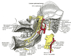
Mandibular division of trigeminal nerve, seen from the middle line. The small figure is an enlarged view of the otic ganglion.
|
|
| Details | |
| From | Trigeminal nerve |
| Identifiers | |
| Latin | Nervus mandibularis |
| MeSH | A08.800.800.120.760.500 |
| Dorlands /Elsevier |
n_05/12566125 |
| TA | A14.2.01.064 |
| FMA | 52996 |
|
Anatomical terms of neuroanatomy
[]
|
|
The mandibular nerve (V3) is the largest of the three divisions of the trigeminal nerve, the fifth cranial nerve (CN V).
The large sensory root emerges from the lateral part of the trigeminal ganglion and exits the cranial cavity through the foramen ovale. The small motor root of the trigeminal nerve passes under the ganglion and through the foramen ovale to unite with the sensory root just outside the skull.
The mandibular nerve immediately passes between tensor veli palatini, which is medial, and lateral pterygoid, which is lateral, and gives off a meningeal branch (nervus spinosus) and the nerve to medial pterygoid from its medial side. The nerve then divides into a small anterior and large posterior trunk.
The anterior division gives off branches to the four main muscles of mastication and a buccal branch which is sensory to the cheek. The posterior division gives off three main sensory branches, the auriculotemporal, lingual and inferior alveolar nerves and motor fibres to supply mylohyoid and the anterior belly of the digastric muscle.
The mandibular nerve gives off the following branches:
The mandibular nerve innervates:
Anterior Division:
(Motor Innervation - Muscles of mastication)
...
Wikipedia
