Trigeminal ganglion
| Trigeminal ganglion | |
|---|---|

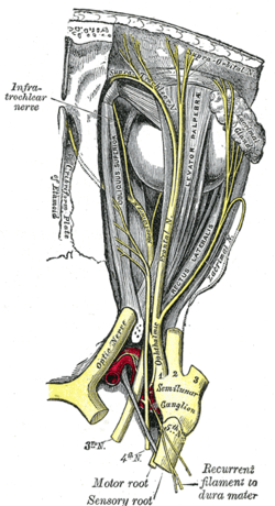
Nerves of the orbit. Seen from above. (Semilunar ganglion visible near bottom.)
|
|

Distribution of the maxillary and mandibular nerves, and the submaxillary ganglion. (Semilunar ganglion visible in upper left.)
|
|
| Details | |
| Identifiers | |
| Latin | ganglion trigeminale, ganglion semilunare (Gasseri) |
| MeSH | A08.340.390.850 |
| Dorlands /Elsevier |
g_02/12385087 |
| TA | A14.2.01.014 |
| FMA | 52618 |
|
Anatomical terms of neuroanatomy
[]
|
|
The trigeminal ganglion (or Gasserian ganglion, or semilunar ganglion, or Gasser's ganglion) is a sensory ganglion of the trigeminal nerve (CN V) that occupies a cavity (Meckel's cave) in the dura mater, covering the trigeminal impression near the apex of the petrous part of the temporal bone.
It is somewhat crescentic in shape, with its convexity directed forward: Medially, it is in relation with the internal carotid artery and the posterior part of the cavernous sinus.
The motor root runs in front of and medial to the sensory root, and passes beneath the ganglion; it leaves the skull through the foramen ovale, and, immediately below this foramen, joins the mandibular nerve.
The greater superficial petrosal nerve lies also underneath the ganglion.
The ganglion receives, on its medial side, filaments from the carotid plexus of the sympathetic.
It gives off minute branches to the tentorium cerebelli, and to the dura mater in the middle fossa of the cranium.
From its convex border, which is directed forward and lateralward, three large nerves proceed, viz., the ophthalmic (V1), maxillary (V2), and mandibular (V3).
The ophthalmic and maxillary consist exclusively of sensory fibers; the mandibular is joined outside the cranium by the motor root.
...
Wikipedia
