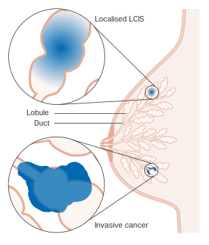
Lobular carcinoma in situ
| Lobular carcinoma in situ | |
|---|---|
 |
|
| Diagram showing localized and invasive LCIS | |
| Classification and external resources | |
| Specialty | oncology |
| ICD-10 | D05.0 |
| ICD-O | M8520/2 |
Lobular carcinoma in situ (LCIS) is a condition caused by unusual cells in the lobules of the breast.
Many do not consider it cancer, but it can indicate an increased risk of future cancer. The national database registrars, however, consider it a malignancy.
Unlike ductal carcinoma in situ (DCIS), LCIS is not associated with calcification, and is typically an incidental finding in a biopsy performed for another reason. LCIS only accounts for about 15% of the in situ (ductal or lobular) breast cancers.
Cells of LCIS and invasive lobular carcinoma have the same histology, appearing as single detached cells, as both have loss of expression of e-cadherin, the transmembrane protein mediating epithelial cell adhesion. LCIS often have the same genetic alterations (such as loss of heterozygosity on chromosome 16q, the locus for the e-cadherin gene) as the adjacent area of invasive carcinoma.
Like the cells of atypical lobular hyperplasia and invasive lobular carcinoma, the abnormal cells of LCIS consist of small cells with oval or round nuclei and small nucleoli detached from each other.Mucin-containing signet-ring cells are commonly seen. LCIS generally leaves the underlying architecture intact and recognisable as lobules. Estrogen and progesterone receptors are present and HER2/neu overexpression is absent in most cases of LCIS.
LCIS may be treated with close clinical follow-up and mammographic screening, tamoxifen or related hormone controlling drugs to reduce the risk of developing cancer, or bilateral prophylactic mastectomy. Some surgeons consider bilateral prophylactic mastectomy to be overly aggressive treatment except for certain high-risk cases.
...
Wikipedia
