Goblet cells
| Goblet cell | |
|---|---|

Schematic illustration of a goblet cell in close up, illustrating different internal structures of the cell.
|
|

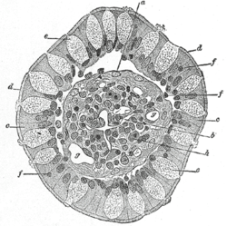
Transverse section of a villus, from the human intestine. X 350.
a. Basement membrane, here somewhat shrunken away from the epithelium. b. Lacteal. c. Columnar epithelium. d. Its striated border. e. Goblet cells. f. Leucocytes in epithelium. f’. Leucocytes below epithelium. g. Blood vessels. h. Muscle cells cut across. |
|
| Details | |
| System | Respiratory system |
| Identifiers | |
| Latin | exocrimohsinocytus caliciformis |
| Code |
TH H3.04.03.0.00009; H3.04.03.0.00016 H3.05.00.0.00006 |
|
Anatomical terminology
[]
|
|
A goblet cell is a glandular, modified simple columnar epithelial cell whose function is to secrete gel-forming mucins, the major components of mucus. The goblet cells mainly use the merocrine method of secretion, secreting vesicles into a duct, but may use apocrine methods, budding off their secretions, when under stress.
The goblet cell is highly polarised with the nucleus and other organelles concentrated at the base of the cell. The remainder of the cell's cytoplasm is occupied by membrane-bound secretory granules containing mucin. The goblet shape is due to the mucus laden granules in the apical part expanding, causing that part of the cell to balloon. The apical plasma membrane projects microvilli to give an increased surface area for secretion.
Goblet cells are found scattered among the epithelial lining of organs, such as the intestinal and respiratory tracts. They are found inside the trachea, bronchi, and larger bronchioles in the respiratory tract, small intestines, the large intestine, and conjunctiva in the upper eyelid. In the conjunctiva goblet cells are a source of mucin in tears and they also secrete different types of mucins onto the ocular surface. In the lacrimal glands, mucus is synthesized by acinar cells instead.
...
Wikipedia
