Fusiform face area
| Fusiform face area | |
|---|---|

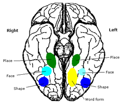
Human brain, bottom view. Fusiform face area shown in bright blue.
|
|

Computer-enhanced fMRI scan of a person who has been asked to look at faces. The image shows increased blood flow in cerebral cortex that recognizes faces (FFA).
|
|
|
Anatomical terminology
[]
|
The fusiform face area (FFA) is a part of the human visual system that, it is speculated, is specialized for facial recognition. It is located in the fusiform gyrus (Brodmann area 37).
The FFA is located in the ventral stream on the ventral surface of the temporal lobe on the lateral side of the fusiform gyrus. It is lateral to the parahippocampal place area. It displays some lateralization, usually being larger in the right hemisphere.
The FFA was discovered and continues to be investigated in humans using positron emission tomography (PET) and functional magnetic resonance imaging (fMRI) studies. Usually, a views images of faces, objects, places, bodies, scrambled faces, scrambled objects, scrambled places, and scrambled bodies. This is called a functional localizer. Comparing the neural response between faces and scrambled faces will reveal areas that are face-responsive, while comparing cortical activation between faces and objects will reveal areas that are face-selective.
The human FFA was first described by Justine Sergent in 1992 and later named by Nancy Kanwisher in 1997 who proposed that the existence of the FFA is evidence for domain specificity in the visual system. More recently, it has been suggested that the FFA processes more than just faces. Some groups, including Isabel Gauthier and others, maintain that the FFA is an area for recognizing fine distinctions between well-known objects. Gauthier et al. tested both car and bird experts, and found some activation in the FFA when car experts were identifying cars and when bird experts were identifying birds. Studies have also suggested that the FFA may be composed of functional clusters that are at a finer spatial scale than prior investigations have measured. Recent evidence also shows that electrical stimulation of these functional clusters selectively distorts face perception, which is causal support for the role of these functional clusters in perceiving the facial image. However, the debate about the functional role of the FFA is ongoing.
...
Wikipedia
