Enolase
| phosphopyruvate hydratase | |||||||||
|---|---|---|---|---|---|---|---|---|---|

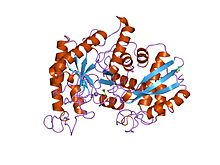
Yeast enolase dimer.
|
|||||||||
| Identifiers | |||||||||
| EC number | 4.2.1.11 | ||||||||
| CAS number | 9014-08-8 | ||||||||
| Databases | |||||||||
| IntEnz | IntEnz view | ||||||||
| BRENDA | BRENDA entry | ||||||||
| ExPASy | NiceZyme view | ||||||||
| KEGG | KEGG entry | ||||||||
| MetaCyc | metabolic pathway | ||||||||
| PRIAM | profile | ||||||||
| PDB structures | RCSB PDB PDBe PDBsum | ||||||||
| Gene Ontology | AmiGO / EGO | ||||||||
|
|||||||||
| Search | |
|---|---|
| PMC | articles |
| PubMed | articles |
| NCBI | proteins |
| Enolase, N-terminal domain | |||||||||
|---|---|---|---|---|---|---|---|---|---|

x-ray structure and catalytic mechanism of lobster enolase
|
|||||||||
| Identifiers | |||||||||
| Symbol | Enolase_N | ||||||||
| Pfam | PF03952 | ||||||||
| Pfam clan | CL0227 | ||||||||
| InterPro | IPR020811 | ||||||||
| PROSITE | PDOC00148 | ||||||||
| SCOP | 1els | ||||||||
| SUPERFAMILY | 1els | ||||||||
|
|||||||||
| Available protein structures: | |
|---|---|
| Pfam | structures |
| PDB | RCSB PDB; PDBe; PDBj |
| PDBsum | structure summary |
| Enolase | |||||||||
|---|---|---|---|---|---|---|---|---|---|

Crystal structure of dimeric beta human enolase ENO3.
|
|||||||||
| Identifiers | |||||||||
| Symbol | Enolase | ||||||||
| Pfam | PF00113 | ||||||||
| InterPro | IPR000941 | ||||||||
| PROSITE | PDOC00148 | ||||||||
|
|||||||||
| Available protein structures: | |
|---|---|
| Pfam | structures |
| PDB | RCSB PDB; PDBe; PDBj |
| PDBsum | structure summary |
Enolase, also known as phosphopyruvate hydratase, is a metalloenzyme responsible for the catalysis of the conversion of 2-phosphoglycerate (2-PG) to phosphoenolpyruvate (PEP), the ninth and penultimate step of glycolysis. The chemical reaction catalyzed by enolase is:
Enolase belongs to the family of lyases, specifically the hydro-lyases, which cleave carbon-oxygen bonds. The systematic name of this enzyme is 2-phospho-D-glycerate hydro-lyase (phosphoenolpyruvate-forming).
The reaction is reversible, depending on environmental concentrations of substrates. The optimum pH for the human enzyme is 6.5. Enolase is present in all tissues and organisms capable of glycolysis or fermentation. The enzyme was discovered by Lohmann and Meyerhof in 1934, and has since been isolated from a variety of sources including human muscle and erythrocytes. In humans, deficiency of ENO1 is linked to hereditary haemolytic anemia while ENO3 deficiency is linked to glycogen storage disease XIII.
In humans there are three subunits of enolase, α, β, and γ, each encoded by a separate gene that can combine to form five different isoenzymes: αα, αβ, αγ, ββ, and γγ. Three of these isoenzymes (all homodimers) are more commonly found in adult human cells than the others:
When present in the same cell, different isozymes readily form heterodimers.
Enolase is a member of the large enolase superfamily. It has a molecular weight of 82,000-100,000 Daltons depending on the isoform. In human alpha enolase, the two subunits are antiparallel in orientation so that Glu20 of one subunit forms an ionic bond with Arg414 of the other subunit. Each subunit has two distinct domains. The smaller N-terminal domain consists of three α-helices and four β-sheets. The larger C-terminal domain starts with two β-sheets followed by two α-helices and ends with a barrel composed of alternating β-sheets and α-helices arranged so that the β-beta sheets are surrounded by the α-helices. The enzyme’s compact, globular structure results from significant hydrophobic interactions between these two domains.
...
Wikipedia
