EGF-like domain
| EGF-like domain | |||||||||
|---|---|---|---|---|---|---|---|---|---|

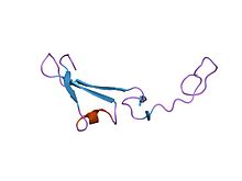
Structure of the epidermal growth factor-like domain of heregulin-alpha.
|
|||||||||
| Identifiers | |||||||||
| Symbol | EGF | ||||||||
| Pfam | PF00008 | ||||||||
| Pfam clan | CL0001 | ||||||||
| InterPro | IPR006209 | ||||||||
| PROSITE | PDOC00021 | ||||||||
| SCOP | 1apo | ||||||||
| SUPERFAMILY | 1apo | ||||||||
| OPM protein | 1dan | ||||||||
| CDD | cd00053 | ||||||||
|
|||||||||
| Available protein structures: | |
|---|---|
| Pfam | structures |
| PDB | RCSB PDB; PDBe; PDBj |
| PDBsum | structure summary |
| EGF-like domain | |||||||||
|---|---|---|---|---|---|---|---|---|---|

crystal structure of the extracellular segment of integrin alphavbeta3
|
|||||||||
| Identifiers | |||||||||
| Symbol | EGF_2 | ||||||||
| Pfam | PF07974 | ||||||||
| Pfam clan | CL0001 | ||||||||
| InterPro | IPR013111 | ||||||||
| CDD | cd00054 | ||||||||
|
|||||||||
| Available protein structures: | |
|---|---|
| Pfam | structures |
| PDB | RCSB PDB; PDBe; PDBj |
| PDBsum | structure summary |
The EGF-like domain is an evolutionary conserved protein domain, which derives its name from the epidermal growth factor where it was first described. It comprises about 30 to 40 amino-acid residues and has been found in a large number of mostly animal proteins. Most occurrences of the EGF-like domain are found in the extracellular domain of membrane-bound proteins or in proteins known to be secreted. An exception to this is the prostaglandin-endoperoxide synthase. The EGF-like domain includes 6 cysteine residues which in the epidermal growth factor have been shown to form 3 disulfide bonds. The structures of 4-disulfide EGF-domains have been solved from the laminin and integrin proteins. The main structure of EGF-like domains is a two-stranded β-sheet followed by a loop to a short C-terminal, two-stranded β-sheet. These two β-sheets are usually denoted as the major (N-terminal) and minor (C-terminal) sheets. EGF-like domains frequently occur in numerous tandem copies in proteins: these repeats typically fold together to form a single, linear solenoid domain block as a functional unit.
Despite the similarities of EGF-like domains, distinct domain subtypes have been identified. The two main proposed types of EGF-like domains are the human EGF-like (hEGF) domain and the complement C1r-like (cEGF) domain, which was first identified in the human complement protease C1r. C1r is a highly specific serine protease initiating the classical pathway of complement activation during immune response. Both the hEGF- and cEGF-like domains contain three disulfides and derive from a common ancestor that carried four disulfides of which one was lost during evolution. Furthermore, cEGF-like domains can be divided in two subtypes (1 and 2) whereas all hEGF-like domains belong to one subtype.
The differentiation of cEGF-like and hEGF-like domains and their subtypes is based on structural features and the connectivity of their disulfide bonds. cEGF- and hEGF-like domains have a distinct shape and orientation of the minor sheet and one C-terminal half-cystine has a different position. The lost cysteines of the common ancestor differ between cEGF- and hEGF-like domains and hence these types differ in their disulfide linkages. The differentiation of cEGF into subtype 1 and 2, which probably occurred after its split from hEGF, is based on different residue numbers between the distinct half-cystines. An N-terminal located calcium binding motif can be found in hEGF- as well as in cEGF-like domains and is therefore not suitable to tell them apart.
...
Wikipedia
