Dilator pupillae
| Iris dilator muscle | |
|---|---|

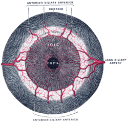
Iris, front view. (Muscle visible but not labeled.)
|
|

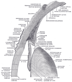
The upper half of a sagittal section through the front of the eyeball. (Iris dilator muscle is NOT labeled and not to be confused with "Radiating fibers" labeled near center, which are part of the ciliary muscle.)
|
|
| Details | |
| Origin | outer margins of iris |
| Insertion | inner margins of iris |
| Nerve | Long ciliary nerves (sympathetics) |
| Actions | dilates pupil |
| Antagonist | iris sphincter muscle |
| Identifiers | |
| Latin | Musculus dilatator pupillae |
| Dorlands /Elsevier |
m_22/12548821 |
| TA | A15.2.03.030 |
| FMA | 49158 |
|
Anatomical terms of muscle
[]
|
|
The iris dilator muscle (pupil dilator muscle, pupillary dilator, radial muscle of iris, radiating fibers), is a smooth muscle of the eye, running radially in the iris and therefore fit as a dilator. The pupillary dilator consists of a spokelike arrangement of modified contractile cells called myoepithelial cells. These cells are stimulated by the sympathetic nervous system. When stimulated, the cells contract, widening the pupil and allowing for more light to pass through the eye.
It is innervated by the sympathetic system, which acts by releasing noradrenaline, which acts on α1-receptors. Thus, when presented with a threatening stimuli that activates the fight-or-flight response, this innervation contracts the muscle and dilates the iris, thus temporarily letting more light reach the retina.
The dilator muscle is innervated more specifically by postganglionic sympathetic nerves arising from the superior cervical ganglion as the sympathetic root of ciliary ganglion. From there, they travel via the internal carotid artery through the carotid canal to foramen lacerum. They then enter the middle cranial fossa above foramen lacerum, travel through the cavernous sinus in the middle cranial fossa and then travel with the ophthalmic artery in the optic canal or on the ophthalmic nerve through the superior orbital fissure. From there, they travel with the nasociliary nerve and then the long ciliary nerve. They then pierce the sclera, travel between sclera and choroid to reach the iris dilator muscle. They will also pass through ciliary ganglion and travel in short ciliary nerves to reach the iris dilator muscle.
...
Wikipedia
