Iris (anatomy)
| Iris | |
|---|---|

Iris is the blue or brown area, with the pupil (the circular black spot) in its center, and surrounded by the white sclera. Overlying cornea is completely transparent so is not visible, except the high-gloss luster it gives the eye. Also pictured are the red blood vessels within the sclera. These structures are easily visible on any person's eyes.
|
|

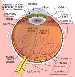
Schematic diagram of the human eye. (Iris labeled at upper right)
|
|
| Details | |
| Precursor | Mesoderm and neural ectoderm |
| Artery | long posterior ciliary arteries |
| Nerve | long ciliary nerves, short ciliary nerves |
| Identifiers | |
| MeSH | A09.371.894.513 |
| TA | A15.2.03.020 |
| FMA | 58235 |
|
Anatomical terminology
[]
|
|
The iris (plural: irides or irises) is a thin, circular structure in the eye, responsible for controlling the diameter and size of the pupil and thus the amount of light reaching the retina. Eye color is defined by that of the iris. In optical terms, the pupil is the eye's aperture, while the iris is the diaphragm that serves as the aperture stop.
The iris consists of two layers: the front pigmented known as a stroma and, beneath the stroma, pigmented epithelial cells.
The stroma connects to a sphincter muscle (sphincter pupillae), which contracts the pupil in a circular motion, and a set of dilator muscles (dilator pupillae) which pull the iris radially to enlarge the pupil, pulling it in folds. The back surface is covered by a heavily pigmented epithelial layer that is two cells thick (the iris pigment epithelium), but the front surface has no epithelium. This anterior surface projects as the dilator muscles. The high pigment content blocks light from passing through the iris to the retina, restricting it to the pupil. The outer edge of the iris, known as the root, is attached to the sclera and the anterior ciliary body. The iris and ciliary body together are known as the anterior uvea. Just in front of the root of the iris is the region referred to as the trabecular meshwork, through which the aqueous humour constantly drains out of the eye, with the result that diseases of the iris often have important effects on intraocular pressure and indirectly on vision. The iris along with the anterior ciliary body provide a secondary pathway for aqueous humour to drain from the eye.
...
Wikipedia
