Choroideremia
| Choroideremia | |
|---|---|
 |
|
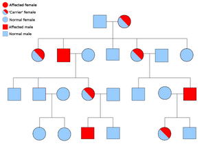
| An example pedigree chart, showing the inheritance of a sex-linked disorder like choroideremia. | |
| Classification and external resources | |
| Specialty | ophthalmology |
| ICD-10 | H31.2 |
| ICD-9-CM | 363.55 |
| OMIM | 303100 |
| DiseasesDB | 2619 |
| MeSH | D015794 |
Choroideremia /kɒˌrɔɪdᵻˈriːmi.ə/ (CHM) is a rare, X-linked recessive form of hereditary retinal degeneration that affects roughly 1 in 50,000 males. The disease causes a gradual loss of vision, starting with childhood night blindness, followed by peripheral vision loss, and progressing to loss of central vision later in life. Progression continues throughout the individual's life, but both the rate of change and the degree of visual loss are variable among those affected, even within the same family.
Choroideremia is caused by a loss-of-function mutation in the CHM gene which encodes Rab escort protein 1 (REP1), a protein involved in lipid modification of Rab proteins. While the complete mechanism of disease is not fully understood, the lack of a functional protein in the retina results in cell death and the gradual deterioration of the choroid, retinal pigment epithelium (RPE), and retinal photoreceptor cells.
As of 2017 there is no treatment for choroideremia, however retinal gene therapy clinical trials have demonstrated a possible treatment.
Since the CHM gene is located on the X chromosome, symptoms are seen almost exclusively in men. While there are a few exceptions, female carriers have a noticeable lack of pigmentation in the RPE but do not experience any symptoms. Female carriers have a 50% chance of having either an affected son or a carrier daughter, while a male with choroideremia will have all carrier daughters and unaffected sons. Even though the disease progression can vary significantly, there are general trends. The first symptom many individuals with choroideremia notice is a significant loss of night vision, which begins in youth.Peripheral vision loss occurs gradually, starting as a ring of vision loss, and continuing on to "tunnel vision" in adulthood. Individuals with choroideremia tend to maintain good visual acuity into their 40s, but eventual lose all sight at some point in the 50-70 age range. A study of 115 individuals with choroideremia found that 84% of patients under the age of 60 had a visual acuity of 20/40 or better, while 33% of patients over 60 years old had a visual acuity of 20/200 or worse. The most severe visual acuity impairment (only being able to count fingers or worse) did not occur until the seventh decade of life. The same study found the rate of visual acuity loss to be about 1 eye chart row per 5 years.
...
Wikipedia
