Retinal pigment epithelium
| Retinal pigment epithelium | |
|---|---|

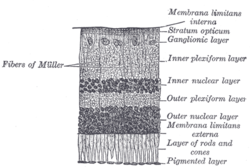
Section of retina. (Pigmented layer labeled at bottom right.)
|
|

Plan of retinal neurons. (Pigmented layer labeled at bottom right.)
|
|
| Details | |
| Identifiers | |
| Latin | Stratum pigmentosum retinae, pars pigmentosa retinae |
| Dorlands /Elsevier |
p_07/{{{DorlandsSuf}}} |
| TA | A15.2.04.008 |
| FMA | 58627 |
|
Anatomical terminology
[]
|
|
The pigmented layer of retina or retinal pigment epithelium (RPE) is the pigmented cell layer just outside the neurosensory retina that nourishes retinal visual cells, and is firmly attached to the underlying choroid and overlying retinal visual cells.
The RPE was known in the 18th and 19th centuries as the pigmentum nigrum, referring to the observation that the RPE is dark (black in many animals, brown in humans) ; and as the tapetum nigrum, referring to the observation that in animals with a tapetum lucidum, in the region of the tapetum lucidum the RPE is not pigmented.
The RPE is composed of a single layer of hexagonal cells that are densely packed with pigment granules.
At the ora serrata, the RPE continues as a membrane passing over the ciliary body and continuing as the back surface of the iris. This generates the fibers of the dilator. Directly beneath this epithelium is the neuroepithelium (i.e., rods and cones) passes jointly with the RPE. Both, combined, are understood to be the ciliary epithelium of the embryo. The front end continuation of the retina is the posterior iris epithelium, which takes on pigment when it enters the iris.
When viewed from the outer surface, these cells are smooth and hexagonal in shape. When seen in section, each cell consists of an outer non-pigmented part containing a large oval nucleus and an inner pigmented portion which extends as a series of straight thread-like processes between the rods, this being especially the case when the eye is exposed to light.
The RPE has several functions, namely, light absorption, epithelial transport, spatial ion buffering, visual cycle, phagocytosis, secretion and immune modulation.
In the eyes of albinos, the cells of this layer contain no pigment. Dysfunction of the RPE is found in age-related macular degeneration and retinitis pigmentosa. RPE are also involved in diabetic retinopathy.
...
Wikipedia
