Cerebral contusion
| Cerebral contusion | |
|---|---|
 |
|
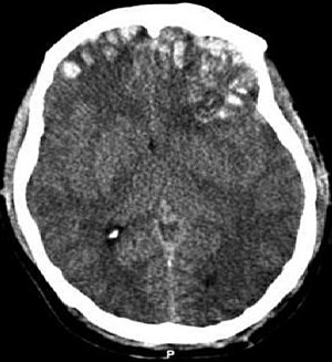
| CT scan showing cerebral contusions, hemorrhage within the hemispheres, subdural hematoma on the left, and skull fractures | |
| Classification and external resources | |
| Specialty | emergency medicine |
| ICD-10 | S06.2, S06.3 |
| ICD-9-CM | 851 |
Cerebral contusion, Latin contusio cerebri, a form of traumatic brain injury, is a bruise of the brain tissue. Like bruises in other tissues, cerebral contusion can be associated with multiple microhemorrhages, small blood vessel leaks into brain tissue. Contusion occurs in 20–30% of severe head injuries. A cerebral laceration is a similar injury except that, according to their respective definitions, the pia-arachnoid membranes are torn over the site of injury in laceration and are not torn in contusion. The injury can cause a decline in mental function in the long term and in the emergency setting may result in brain herniation, a life-threatening condition in which parts of the brain are squeezed past parts of the skull. Thus treatment aims to prevent dangerous rises in intracranial pressure, the pressure within the skull.
Contusions are likely to heal on their own without medical intervention.
The symptoms of a cerebral contusion (bruising on the brain) depend on the severity of the injury, ranging from minor to severe. Individuals may experience a headache; confusion; sleepiness; dizziness; loss of consciousness; nausea and vomiting; seizures; and difficulty with coordination and movement. They may also have difficulty with memory, vision, speech, hearing, managing emotions, and thinking. Signs depend on the contusion's location in the brain.
Often caused by a blow to the head, contusions commonly occur in coup or contre-coup injuries. In coup injuries, the brain is injured directly under the area of impact, while in contrecoup injuries it is injured on the side opposite the impact.
Contusions occur primarily in the cortical tissue, especially under the site of impact or in areas of the brain located near sharp ridges on the inside of the skull. The brain may be contused when it collides with bony protuberances on the inside surface of the skull. The protuberances are located on the inside of the skull under the frontal and temporal lobes and on the roof of the ocular orbit. Thus, the tips of the frontal and temporal lobes located near the bony ridges in the skull are areas where contusions frequently occur and are most severe. For this reason, attention, emotional and memory problems, which are associated with damage to frontal and temporal lobes, are much more common in head trauma survivors than are syndromes associated with damage to other areas of the brain.
...
Wikipedia
