Arrestins
| S-antigen; retina and pineal gland (arrestin) | |
|---|---|

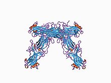
Crystallographic structure of the bovine arrestin-S.
|
|
| Identifiers | |
| Symbol | SAG |
| Alt. symbols | arrestin-1 |
| Entrez | 6295 |
| HUGO | 10521 |
| OMIM | 181031 |
| RefSeq | NM_000541 |
| UniProt | P10523 |
| Other data | |
| Locus | Chr. 2 q37.1 |
| arrestin beta 1 | |
|---|---|
| Identifiers | |
| Symbol | ARRB1 |
| Alt. symbols | ARR1, arrestin-2 |
| Entrez | 408 |
| HUGO | 711 |
| OMIM | 107940 |
| RefSeq | NM_004041 |
| UniProt | P49407 |
| Other data | |
| Locus | Chr. 11 q13 |
| arrestin beta 2 | |
|---|---|
| Identifiers | |
| Symbol | ARRB2 |
| Alt. symbols | ARR2, arrestin-3 |
| Entrez | 409 |
| HUGO | 712 |
| OMIM | 107941 |
| RefSeq | NM_004313 |
| UniProt | P32121 |
| Other data | |
| Locus | Chr. 17 p13 |
| arrestin 3, retinal (X-arrestin) | |
|---|---|
| Identifiers | |
| Symbol | ARR3 |
| Alt. symbols | ARRX, arrestin-4 |
| Entrez | 407 |
| HUGO | 710 |
| OMIM | 301770 |
| RefSeq | NM_004312 |
| UniProt | P36575 |
| Other data | |
| Locus | Chr. X q |
| Arrestin (or S-antigen), N-terminal domain | |||||||||
|---|---|---|---|---|---|---|---|---|---|

Structure of arrestin from bovine rod outer segments.
|
|||||||||
| Identifiers | |||||||||
| Symbol | Arrestin_N | ||||||||
| Pfam | PF00339 | ||||||||
| Pfam clan | CL0135 | ||||||||
| InterPro | IPR011021 | ||||||||
| PROSITE | PDOC00267 | ||||||||
| SCOP | 1cf1 | ||||||||
| SUPERFAMILY | 1cf1 | ||||||||
|
|||||||||
| Available protein structures: | |
|---|---|
| Pfam | structures |
| PDB | RCSB PDB; PDBe; PDBj |
| PDBsum | structure summary |
| Arrestin (or S-antigen), C-terminal domain | |||||||||
|---|---|---|---|---|---|---|---|---|---|

Structure of bovine beta-arrestin.
|
|||||||||
| Identifiers | |||||||||
| Symbol | Arrestin_C | ||||||||
| Pfam | PF02752 | ||||||||
| Pfam clan | CL0135 | ||||||||
| InterPro | IPR011022 | ||||||||
| SCOP | 1cf1 | ||||||||
| SUPERFAMILY | 1cf1 | ||||||||
|
|||||||||
| Available protein structures: | |
|---|---|
| Pfam | structures |
| PDB | RCSB PDB; PDBe; PDBj |
| PDBsum | structure summary |
Arrestins are a small family of proteins important for regulating signal transduction at G protein-coupled receptors. Arrestins were first discovered as a part of a conserved two-step mechanism for regulating the activity of G protein-coupled receptors (GPCRs) in the visual rhodopsin system by Hermann Kühn and co-workers and in the β-adrenergic system by Martin J. Lohse and co-workers.
In response to a stimulus, GPCRs activate heterotrimeric G proteins. In order to turn off this response, or adapt to a persistent stimulus, active receptors need to be desensitized. The first step is phosphorylation by a class of serine/threonine kinases called G protein coupled receptor kinases (GRKs). GRK phosphorylation specifically prepares the activated receptor for arrestin binding. Arrestin binding to the receptor blocks further G protein-mediated signaling and targets receptors for internalization, and redirects signaling to alternative G protein-independent pathways, such as β-arrestin signaling. In addition to GPCRs, arrestins bind to other classes of cell surface receptors and a variety of other signaling proteins.
Mammals express four arrestin subtypes and each arrestin subtype is known by multiple aliases. The systematic arrestin name (1-4) plus the most widely used aliases for each arrestin subtype are listed in bold below:
Fish and other vertebrates appear to have only three arrestins: no equivalent of arrestin-2, which is the most abundant non-visual subtype in mammals, was cloned so far. The proto-chordate C. intestinalis (sea squirt) has only one arrestin, which serves as visual in its mobile larva with highly developed eyes, and becomes generic non-visual in the blind sessile adult. Conserved positions of multiple introns in its gene and those of our arrestin subtypes suggest that they all evolved from this ancestral arrestin. Lower invertebrates, such as roundworm C. elegans, also have only one arrestin. Insects have arr1 and arr2, originally termed “visual arrestins” because they are expressed in photoreceptors, and one non-visual subtype (kurtz in Drosophila). Later arr1 and arr2 were found to play an important role in olfactory neurons and renamed “sensory”. Fungi have distant arrestin relatives involved in pH sensing.
...
Wikipedia
