Wallerian degeneration
| Nerve injury | |
|---|---|
 |
|
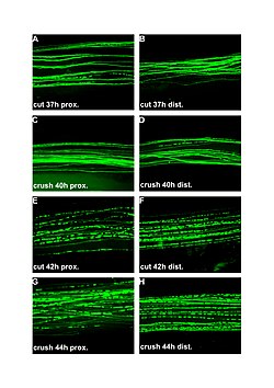
| Fluorescent micrographs (100x) of Wallerian degeneration in cut and crushed peripheral nerves. Left column is proximal to the injury, right is distal. A and B: 37 hours post cut. C and D: 40 hours post crush. E and F: 42 hours post cut. G and H: 44 hours post crush. | |
| Classification and external resources | |
| MeSH | D014855 |
Wallerian degeneration is a process that results when a nerve fiber is cut or crushed, in which the part of the axon separated from the neuron's cell body degenerates distal to the injury. This is also known as anterograde or orthograde degeneration. A related process known as 'Wallerian-like degeneration' occurs in many neurodegenerative diseases, especially those where axonal transport is impaired.Primary culture studies suggest that a failure to deliver sufficient quantities of the essential axonal protein NMNAT2 is a key initiating event.
Wallerian degeneration occurs after axonal injury in both the peripheral nervous system (PNS) and central nervous system (CNS). It occurs in the axon stump distal to a site of injury and usually begins within 24–36 hours of a lesion. Prior to degeneration, distal axon stumps tend to remain electrically excitable. After injury, the axonal skeleton disintegrates, and the axonal membrane breaks apart. The axonal degeneration is followed by degradation of the myelin sheath and infiltration by macrophages. The macrophages, accompanied by Schwann cells, serve to clear the debris from the degeneration.
The nerve fiber's neurolemma does not degenerate and remains as a hollow tube. Within 4 days of the injury, the distal end of the portion of the nerve fiber proximal to the lesion sends out sprouts towards those tubes and these sprouts are attracted by growth factors produced by Schwann cells in the tubes. If a sprout reaches the tube, it grows into it and advances about 1 mm per day, eventually reaching and reinnervating the target tissue. If the sprouts cannot reach the tube, for instance because the gap is too wide or scar tissue has formed, surgery can help to guide the sprouts into the tubes. This regeneration is much slower in the spinal cord than in PNS. The crucial difference is that in the CNS, including in the spinal cord, myelin sheaths are produced by oligodendrocytes and not by Schwann cell.
...
Wikipedia
