Thioredoxin reductase
| Thioredoxin-disulfide reductase | |||||||||
|---|---|---|---|---|---|---|---|---|---|

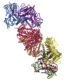
Crystal structure of human thioredoxin reductase 1; rendering based on PDB: 2OHV.
|
|||||||||
| Identifiers | |||||||||
| EC number | 1.8.1.9 | ||||||||
| CAS number | 9074-14-0 | ||||||||
| Databases | |||||||||
| IntEnz | IntEnz view | ||||||||
| BRENDA | BRENDA entry | ||||||||
| ExPASy | NiceZyme view | ||||||||
| KEGG | KEGG entry | ||||||||
| MetaCyc | metabolic pathway | ||||||||
| PRIAM | profile | ||||||||
| PDB structures | RCSB PDB PDBe PDBsum | ||||||||
| Gene Ontology | AmiGO / EGO | ||||||||
|
|||||||||
| Search | |
|---|---|
| PMC | articles |
| PubMed | articles |
| NCBI | proteins |
| thioredoxin reductase 1 | |
|---|---|
| Identifiers | |
| Symbol | TXNRD1 |
| Entrez | 7296 |
| HUGO | 12437 |
| OMIM | 601112 |
| RefSeq | NM_003330 |
| UniProt | Q16881 |
| Other data | |
| EC number | 1.8.1.9 |
| Locus | Chr. 12 q23-q24.1 |
| thioredoxin reductase 2 | |
|---|---|
| Identifiers | |
| Symbol | TXNRD2 |
| Entrez | 10587 |
| HUGO | 18155 |
| OMIM | 606448 |
| RefSeq | NM_006440 |
| UniProt | Q9NNW7 |
| Other data | |
| EC number | 1.8.1.9 |
| Locus | Chr. 22 q11.21 |
| thioredoxin reductase 3 | |
|---|---|
| Identifiers | |
| Symbol | TXNRD3 |
| Entrez | 114112 |
| HUGO | 20667 |
| OMIM | 606235 |
| RefSeq | XM_051264 |
| UniProt | Q86VQ6 |
| Other data | |
| EC number | 1.8.1.9 |
| Locus | Chr. 3 p13-q13.33 |
Thioredoxin reductases (TR, TrxR) (EC 1.8.1.9) are the only known enzymes to reduce thioredoxin (Trx). Two classes of thioredoxin reductase have been identified: one class in bacteria and some eukaryotes and one in animals. Both classes are flavoproteins which function as homodimers. Each monomer contains a FAD prosthetic group, a NADPH binding domain, and an active site containing a redox-active disulfide bond.
Thioredoxin reductase is the only enzyme known to catalyze the reduction of thioredoxin and hence is a central component in the thioredoxin system. Together with thioredoxin (Trx) and NADPH this system's most general description is as a method of forming reduced disulfide bonds in cells. Electrons are taken from NADPH via TrxR and are transferred to the active site of Trx, which goes on to reduce protein disulfides or other substrates. The Trx system exists in all living cells and has an evolutionary history tied to DNA as a genetic material, defense against oxidative damage due to oxygen metabolism, and redox signaling using molecules like hydrogen peroxide and nitric oxide.
Two classes of thioredoxin reductase have evolved independently:
These two classes of TrxR have only ~20% sequence identity in the section of primary sequence where they can be reliably aligned. The net reaction of both classes of TrxR is identical but the mechanism of action of each is distinct.
Humans express three thioredoxin reductase isozymes: TrxR1 (cytosolic), TrxR2 (mitochondrial), TrxR3 (testis specific). Each isozyme is encoded by a separate gene:
E. coli thioredoxin reductase structure: In E. coli ThxR there are two binding domains, one for FAD and another for NADPH. The connection between these two domains is a two-stranded anti-parallel β-sheet. Each domain individually is very similar to the analogous domains in glutathione reductase, and lipoamide dehydrogenase but they relative orientation of these domains in ThxR is rotated by 66 degrees. This becomes significant in the enzyme mechanism of action which is described below. ThxR homo-dimerizes with the interface between the two monomers formed by three alpha-helices and two loops. Each monomer can separately bind a molecule of thioredoxin.
...
Wikipedia
