Supracondylar fracture
| Supracondylar humerus fracture | |
|---|---|
| Classification and external resources | |

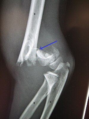
An elbow X-ray showing a displaced supracondylar fracture in a young child
|
|
| ICD-10 | S42.4 |
| AO | 13-A2.3 |
| eMedicine | 1269576 |
A supracondylar humerus fracture is a fracture of the distal humerus just above the elbow joint. The fracture is usually transverse or oblique and above the medial and lateral condyles and epicondyles. This fracture pattern is relatively rare in adults, but is the most common type of elbow fracture in children. In children, many of these fractures are non-displaced and can be treated with casting. Some are angulated or displaced and are best treated with surgery. In children, most of these fractures can be treated effectively with expectation for full recovery. Some of these injuries can be complicated by poor healing or by associated blood vessel or nerve injuries with serious complications.
The fracture is caused by a fall on an outstretched hand (FOOSH) in 70% of cases. As the hand hits the ground, the elbow is hyperextended, resulting in a fracture of the distal humerus above the condyles. With hyperextension, the olecranon process of the ulna is forced against the weaker, immature metaphyseal bone of the distal humerus, producing the typical extension-type supracondylar fracture”. A flexion type fracture can result from a direct blow to the posterior aspect of the elbow when the elbow is in a flexed position. This causes the distal condylar fragment to displace in an anterior direction.
Supracondylar humerus fractures typically result from a fall on to an outstretched arm, usually leading to a forced hyperextension of the elbow. Typically, this is an isolated injury to the elbow (no injuries elsewhere). Children with this injury present with pain and swelling about the elbow. Motion at the elbow and at the wrist make the pain worse. With mild or moderate fracture displacement, there may be deformity at the elbow.
Diagnosis is confirmed by x-ray imaging. Displaced fractures are readily apparent. A non-displaced fracture can be difficult to identify and a fracture line may not be visible on the X-rays. However, the presence of a joint effusion is highly suggestive of a non-displaced fracture. Bleeding from the fracture expands the joint capsule and is visualized on the lateral view as a darker area anteriorly and posteriorly, and is known as the sail sign. Depending on the child's age, parts of the bone will still be developing and if not yet calcified, will not show up on the X-rays. At times, X-rays of the opposite elbow may be obtained for comparison. There are landmarks on the X-rays that can be used to assess displacement.
...
Wikipedia
