Projectional radiography
| Projectional radiography | |
|---|---|
| Intervention | |

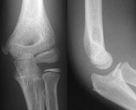
AP and Lateral Elbow X-Ray
|
|
| ICD-10-PCS | B?0 |
| ICD-9-CM | 87 |
| OPS-301 code | 3-10...3-13 |
Projectional radiography is the practice of producing two-dimensional images using x-ray radiation. Radiographic exams are typically performed by radiographers, trained, licensed medical professionals. Projectional radiography is the cornerstone of modern medical imaging, and can be used to image almost every part of the human body. Mammography, DXA and dental radiography are specialized variants of projectional radiography. Computed tomography also uses x-radiation, but its different image acquisition parameters allow for images to be acquired in the axial plane.
Projectional radiography relies on the characteristics of x-ray radiation (quantity and quality of the beam) and knowledge of how it interacts with human tissue to create diagnostic images. X-rays are a form of ionizing radiation, meaning it has sufficient energy to potentially remove electrons from an atom, thus giving it a charge and making it an ion. Ionizing radiation has sufficient energy to penetrate human tissue.
When an exposure is made, x-ray radiation exits the tube as what is known as the primary beam. When the primary beam passes through the body, some of the radiation is absorbed in a process known as attenuation. Anatomy that is denser has a higher rate of attenuation than anatomy that is less dense, so bone will absorb more x-rays than soft tissue. What remains of the primary beam after attenuation is known as the remnant beam. The remnant beam is responsible for exposing the image receptor. Areas on the image receptor that receive the most radiation (portions of the remnant beam experiencing the least attenuation) will be more heavily exposed, and therefore will be processed as being darker. Conversely, areas on the image receptor that receive the least radiation (portions of the remnant beam experience the most attenuation) will be less exposed and will be processed as being lighter. This is why bone, which is very dense, process as being ‘white’ on radio graphs, and the lungs, which contain mostly air and is the least dense, shows up as ‘black’.
Radiographic density is the measure of overall darkening of the image. Density is a logarithmic unit that describes the ratio between light hitting the film and light being transmitted through the film. A higher radiographic density represents more opaque areas of the film, and lower density more transparent areas of the film.
With digital imaging, however, density may be referred to as brightness. The brightness of the radiograph in digital imaging is determined by computer software and the monitor on which the image is being viewed.
...
Wikipedia
