Submucosa
| Submucosa | |
|---|---|

|
|

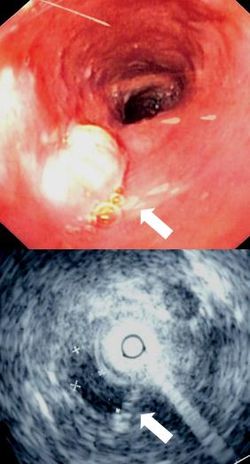
Endoscopy and radial endoscopic ultrasound images of submucosal tumour in mid-esophagus. The submucosa is seen as a dark ring on the ultrasound image.
|
|
| Details | |
| Identifiers | |
| Latin | tela submucosa |
| TA | A05.3.01.028 |
| FMA | 85391 55072, 85391 |
|
Anatomical terminology
[]
|
|
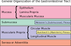
The submucosa (or tela submucosa) is a thin layer of tissue in various organs of the gastrointestinal, respiratory, and genitourinary tracts. It is the layer of dense irregular connective tissue that supports the mucosa (mucous membrane) and joins it to the muscular layer, the bulk of overlying smooth muscle (fibers running circularly within layer of longitudinal muscle).
The submucosa ( + mucosa) is to a mucous membrane what the subserosa ( + serosa) is to a serous membrane.
Blood vessels, lymphatic vessels, and nerves (all supplying the mucosa) will run through here. Tiny parasympathetic ganglia are scattered around forming the submucosal plexus (or "Meissner's plexus") where preganglionic parasympathetic neurons synapse with postganglionic nerve fibers that supply the muscularis mucosae. Histologically, the wall of the alimentary canal shows four distinct layers (from the lumen moving out): mucosa, submucosa, muscularis externa, and a serosa or adventitia.
Identification of the submucosa plays an important role in diagnostic and therapeutic endoscopy, where special fibre-optic cameras are used to perform procedures on the gastrointestinal tract. Abnormalities of the submucosa, such as gastrointestinal stromal tumors, usually show integrity of the mucosal surface.
...
Wikipedia
