Auerbach's plexus
| Myenteric plexus | |
|---|---|

The myenteric plexus from the rabbit. X 50.
|
|
 |
|
| Details | |
| Identifiers | |
| Latin | Plexus myentericus, plexus Auerbachi |
| MeSH | A08.800.050.050.500 |
| TA | A14.3.03.041 |
| FMA | 63252 |
|
Anatomical terms of neuroanatomy
[]
|
|
The myenteric plexus (or Auerbach's plexus) provides motor innervation to both layers of the muscular layer of the gut, having both parasympathetic and sympathetic input (although present ganglionar cell bodies belong to parasympathetic innervation, fibers from sympathetic innervation also reach the plexus), whereas the submucous plexus has only parasympathetic fibers and provides secretomotor innervation to the mucosa nearest the lumen of the gut.
It arises from cells in the vagal trigone also known as the nucleus ala cinerea, the parasympathetic nucleus of origin for the tenth cranial nerve (vagus nerve), located in the medulla oblongata. The fibers are carried by both the anterior and posterior vagal nerves. The myenteric plexus is the major nerve supply to the gastrointestinal tract and controls GI tract motility.
According to preclinical studies, 30% of myenteric plexus' neurons are enteric sensory neurons, thus Auerbach's plexus has also a sensory component.
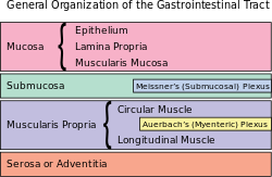
A part of the enteric nervous system, the myenteric plexus exists between the longitudinal and circular layers of muscularis externa in the gastrointestinal tract. It is found in the muscles of the esophagus, stomach, and intestine.
The ganglia have properties similar to the central nervous system (CNS). These properties include presence of glia, interneurons, a small extracellular space, dense synaptic neuropil, isolation from blood vessels, multiple synaptic mechanisms and multiple neurotransmitters.
The myenteric plexus originates in the medulla oblongata as a collection of neurons from the ventral part of the brain stem. The vagus nerve then carries the axons to their destination in the gastrointestinal tract.
They contain Dogiel cells.
The myenteric plexus functions as a part of the enteric nervous system (digestive system). The enteric nervous system can and does function autonomously, but normal digestive function requires communication links between this intrinsic system and the central nervous system. The ENS contains sensory receptors, primary afferent neurons, interneurons, and motor neurons. The events that are controlled, at least in part, by the ENS are multiple and include motor activity, secretion, absorption, blood flow, and interaction with other organs such as the gallbladder or pancreas. These links take the form of parasympathetic and sympathetic fibers that connect either the central and enteric nervous systems or connect the central nervous system directly with the digestive tract. Through these cross connections, the gut can provide sensory information to the CNS, and the CNS can affect gastrointestinal function. Connection to the central nervous system also means that signals from outside of the digestive system can be relayed to the digestive system: for instance, the sight of appealing food stimulates secretion in the stomach.
...
Wikipedia
