Squalene synthase
| Squalene synthase | |||||||||
|---|---|---|---|---|---|---|---|---|---|

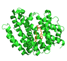
Human Squalene synthase in complex with inhibitor. PDB 3q30
|
|||||||||
| Identifiers | |||||||||
| EC number | 2.5.1.21 | ||||||||
| CAS number | 9077-14-9 | ||||||||
| Databases | |||||||||
| IntEnz | IntEnz view | ||||||||
| BRENDA | BRENDA entry | ||||||||
| ExPASy | NiceZyme view | ||||||||
| KEGG | KEGG entry | ||||||||
| MetaCyc | metabolic pathway | ||||||||
| PRIAM | profile | ||||||||
| PDB structures | RCSB PDB PDBe PDBsum | ||||||||
| Gene Ontology | AmiGO / EGO | ||||||||
|
|||||||||
| Search | |
|---|---|
| PMC | articles |
| PubMed | articles |
| NCBI | proteins |
| farnesyl-diphosphate farnesyltransferase 1 | |
|---|---|
| Identifiers | |
| Symbol | FDFT1 |
| Entrez | 2222 |
| HUGO | 3629 |
| OMIM | 184420 |
| RefSeq | NM_004462 |
| UniProt | P37268 |
| Other data | |
| EC number | 2.5.1.21 |
| Locus | Chr. 8 p23.1-p22 |
Squalene synthase (SQS) or farnesyl-diphosphate:farnesyl-diphosphate farnesyl transferase is an enzyme localized to the membrane of the endoplasmic reticulum. SQS participates in the isoprenoid biosynthetic pathway, catalyzing a two-step reaction in which two identical molecules of farnesyl pyrophosphate (FPP) are converted into squalene, with the consumption of NADPH.Catalysis by SQS is the first committed step in sterol synthesis, since the squalene produced is converted exclusively into various sterols, such as cholesterol, via a complex, multi-step pathway. SQS belongs to squalene/phytoene synthase family of proteins.
Squalene synthase has been characterized in animals, plants, and yeast. In terms of structure and mechanics, squalene synthase closely resembles phytoene synthase (PHS), another prenyltransferase. PHS serves a similar role to SQS in plants and bacteria, catalyzing the synthesis of phytoene, a precursor of carotenoid compounds.
Squalene synthase (SQS) is localized exclusively to the membrane of the endoplasmic reticulum (ER). SQS is anchored to the membrane by a short C-terminal membrane-spanning domain. The N-terminal catalytic domain of the enzyme protrudes into the cytosol, where the soluble substrates are bound. Mammalian forms of SQS are approximately 47kDa and consist of ~416 amino acids. The crystal structure of human SQS was determined in 2000, and revealed that the protein was composed entirely of α-helices. The enzyme is folded into a single domain, characterized by a large central channel. The active sites of both of the two half-reactions catalyzed by SQS are located within this channel. One end of the channel is open to the cytosol, whereas the other end forms a hydrophobic pocket. SQS contains two conserved aspartate-rich sequences, which are believed to participate directly in the catalytic mechanism. These aspartate-rich motifs are one of several conserved structural features in class I isoprenoid biosynthetic enzymes, although these enzymes do not share sequence homology.
...
Wikipedia
