Spindle cell
| Spindle neuron | |
|---|---|

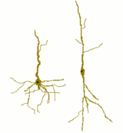
Cartoon of a spindle cell (right) compared to a normal pyramidal cell (left).
|
|
| Details | |
| Location | Anterior cingulate cortex and Fronto-insular cortex |
| Morphology | Unique spindle-shaped projection neuron |
| Function | Global firing rate regulation and regulation of emotional state |
| Presynaptic connections | Local input to ACC and FI |
| Postsynaptic connections | Frontal and temporal cortex. |
|
Anatomical terminology
[]
|
|
Spindle neurons, also called von Economo neurons (VENs), are a specific class of neurons that are characterized by a large spindle-shaped soma (or body), gradually tapering into a single apical axon in one direction, with only a single dendrite facing opposite. Other neurons tend to have many dendrites, and the polar-shaped morphology of spindle neurons is unique. A neuron's dendrites receive signals, and its axon sends them.
Spindle neurons are found in two very restricted regions in the brains of hominids—the family of species comprising humans and other great apes—the anterior cingulate cortex (ACC) and the fronto-insular cortex (FI). Recently they have been discovered in the dorsolateral prefrontal cortex of humans. Spindle cells are also found in the brains of the humpback whales, fin whales, killer whales, sperm whales,bottlenose dolphin, Risso’s dolphin, beluga whales,African and Asian elephants, and to a lesser extent in macaque monkeys and raccoons. The appearance of spindle neurons in distantly related clades suggests that they represent convergent evolution, specifically an adaptation to larger brains.
...
Wikipedia
