Pupils
| Pupil | |
|---|---|

|
|

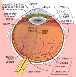
Schematic diagram of the human eye.
|
|
| Details | |
| Identifiers | |
| Latin | Pupilla (Plural: Pupillae) |
| TA | A15.2.03.028 |
| FMA | 58252 |
|
Anatomical terminology
[]
|
|
The pupil is a hole located in the centre of the iris of the eye that allows light to strike the retina. It appears black because light rays entering the pupil are either absorbed by the tissues inside the eye directly, or absorbed after diffuse reflections within the eye that mostly miss exiting the narrow pupil.
In humans the pupil is round, but other species, such as some cats, have vertical slit pupils, goats have horizontally oriented pupils, and some catfish have annular types. In optical terms, the anatomical pupil is the eye's aperture and the iris is the aperture stop. The image of the pupil as seen from outside the eye is the entrance pupil, which does not exactly correspond to the location and size of the physical pupil because it is magnified by the cornea. On the inner edge lies a prominent structure, the collarette, marking the junction of the embryonic pupillary membrane covering the embryonic pupil.
The iris is a contractile structure, consisting mainly of smooth muscle, surrounding the pupil. Light enters the eye through the pupil, and the iris regulates the amount of light by controlling the size of the pupil. The iris contains two groups of smooth muscles; a circular group called the sphincter pupillae, and a radial group called the dilator pupillae. When the sphincter pupillae contract, the iris decreases or constricts the size of the pupil. The dilator pupillae, innervated by sympathetic nerves from the superior cervical ganglion, cause the pupil to dilate when they contract. These muscles are sometimes referred to as intrinsic eye muscles. The sensory pathway (rod or cone, bipolar, ganglion) is linked with its counterpart in the other eye by a partial crossover of each eye's fibers. This causes the effect in one eye to carry over to the other. If the drug pilocarpine is administered, the pupils will constrict and accommodation is increased due to the parasympathetic action on the circular muscle fibers, conversely, atropine will cause paralysis of accommodation (cycloplegia) and dilation of the pupil.
...
Wikipedia
