Primary auditory cortex
| Primary auditory cortex | |
|---|---|

|
|

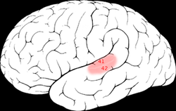
The primary auditory cortex is highlighted in pink and interacts with the other areas highlighted above
|
|
| Details | |
| Identifiers | |
| Latin | Cortex auditivus primus |
| NeuroNames | ancil-428 |
| FMA | 226221 |
|
Anatomical terms of neuroanatomy
[]
|
|
The primary auditory cortex is the part of the temporal lobe that processes auditory information in humans and other vertebrates. It is a part of the auditory system, performing basic and higher functions in hearing.
It is located bilaterally, roughly at the upper sides of the temporal lobes – in humans on the superior temporal plane, within the lateral fissure and comprising parts of Heschl's gyrus and the superior temporal gyrus, including planum polare and planum temporale (roughly Brodmann areas 41, 42, and partially 22). Unilateral destruction results in slight hearing loss, whereas bilateral destruction results in cortical deafness.
The auditory cortex was previously subdivided into primary (A1) and secondary (A2) projection areas and further association areas. The modern divisions of the auditory cortex are the core (which includes A1), the belt, and the parabelt. The belt is the area immediately surrounding the core; the parabelt is adjacent to the lateral side of the belt.
Besides receiving input from the ears via lower parts of the auditory system, it also transmits signals back to these areas and is interconnected with other parts of the cerebral cortex.
Data about the auditory cortex has been obtained through studies in rodents, cats, macaques, and other animals. In humans, the structure and function of the auditory cortex has been studied using functional magnetic resonance imaging (fMRI), electroencephalography (EEG), and electrocorticography.
Like many areas in the neocortex, the functional properties of the adult primary auditory cortex (A1) are highly dependent on the sounds encountered early in life. This has been best studied using animal models, especially cats and rats. In the rat, exposure to a single frequency during postnatal day (P) 11 to 13 can cause a 2-fold expansion in the representation of that frequency in A1. Importantly, the change is persistent, in that it lasts throughout the animal's life, and specific, in that the same exposure outside of that period causes no lasting change in the tonotopy of A1.
...
Wikipedia
