Pineal organ
| Pineal gland | |
|---|---|

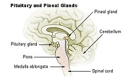
Diagram of pituitary and pineal glands in the human brain
|
|
| Details | |
| Precursor | Neural ectoderm, roof of diencephalon |
| Artery | posterior cerebral artery |
| Identifiers | |
| Latin | glandula pinealis |
| MeSH | Pineal+gland |
| NeuroLex ID | Pineal body |
| Dorlands /Elsevier |
g_06/12392585 |
| TA | A11.2.00.001 |
| FMA | 62033 |
|
Anatomical terms of neuroanatomy
[]
|
|
The pineal gland, also known as the pineal body, conarium or epiphysis cerebri, is a small endocrine gland in the vertebrate brain. The shape of the gland resembles a pine cone, hence its name. The pineal gland is located in the epithalamus, near the center of the brain, between the two hemispheres, tucked in a groove where the two halves of the thalamus join. The pineal gland produces melatonin (not to be confused with melanin), a serotonin derived hormone which modulates sleep patterns in both circadian and seasonal cycles.
Nearly all vertebrate species possess a pineal gland. The most important exception is the hagfish, which is often thought of as the most primitive extant vertebrate. Even in the hagfish, however, there may be a "pineal equivalent" structure in the dorsal diencephalon. The lancelet Branchiostoma lanceolatum, the nearest existing relative to vertebrates, also lacks a recognizable pineal gland. The lamprey (considered almost as primitive as the hagfish), however, does possess one. A few more developed vertebrates lost pineal glands over the course of their evolution.
The results of various scientific research in evolutionary biology, comparative neuroanatomy and neurophysiology, have explained the phylogeny of the pineal gland in different vertebrate species. From the point of view of biological evolution, the pineal gland represents a kind of atrophied photoreceptor. In the epithalamus of some species of amphibians and reptiles, it is linked to a light-sensing organ, known as the parietal eye, which is also called the pineal eye or third eye.
...
Wikipedia
