Paracoccidioides
| Paracoccidioides brasiliensis | |
|---|---|
 |
|
| Scientific classification | |
| Kingdom: | Fungi |
| Division: | Ascomycota |
| Class: | Eurotiomycetes |
| Order: | Onygenales |
| Family: | Ajellomycetaceae |
| Genus: | Paracoccidioides |
| Species: | P. brasiliensis |
| Binomial name | |
|
Paracoccidioides brasiliensis (Splend.) F.P.Almeida (1930) |
|
| Synonyms | |
|
Zymonema brasiliensis Splend. (1912) |
|
Zymonema brasiliensis Splend. (1912)
Coccidioides brasiliensis F.P.Almeida (1929)
Paracoccidioides brasiliensis is a dimorphic fungus and the causative agent of the disease paracoccidioidomycosis. The fungus has been affiliated with the family Ajellomycetaceae (division Ascomycota) although a sexual state or teleomorph has not yet been found.
Paracoccidioides brasiliensis was first discovered by Adolfo Lutz in 1908 in Brazil. Although Lutz did not suggest a name for the disease caused by this fungus, he made note of structures he called “pseudococcidica” together with mycelium in cultures grown at 25 °C. In 1912, Alfonse Splendore proposed the name Zymonema brasiliense and described the features of the fungus in culture. Finally in 1930, Floriano de Almeida created the genus Paracoccidioides to accommodate the species, noting its distinction from Coccidioides immitis.
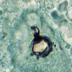
Paracoccidioides brasiliensis is a nonphotosynthetic eukaryote with a rigid cell wall and organelles very similar to those of higher eukaryotes. Being a dimorphic fungus, it has the ability to grow an oval yeast-like form at 37 °C and an elongated mycelial form produced at room temperature. The mycelial and yeast phases differ in their morphology, biochemistry, and ultrastructure. The yeast form contains large amounts of α-(1,3)-linked glucan. The chitin content of the mycelial form is greater than that of the yeast form, but the lipid content of both phases is comparable. The yeast reproduces by asexual budding, where daughter cells are borne asynchronously at multiple, random positions across the cell surface. Buds begin by layers of cell wall increasing in optical density at a point that eventually gives rise to the daughter cell. Once the bud has expanded, a cleavage plane develops between the nascent cell and the mother cell. Following dehiscence, the bud scar disappears. In tissue, budding occurs inside the granulomatous center of the disease lesion, as visualized by hematoxylin and eosin (H&E) staining of histologic sections. Nonbudding cells measure 5–15 µm in diameter, whereas those with multiple spherical buds measure from 10–20 µm in diameter. In electron microscopy, cells with multiple buds have been found to have peripherally located nuclei and cytoplasm surrounding a large central vacuole. In the tissue form of P. brasiliensis, yeast cells are larger with thinner walls and a narrower bud base than those of the related dimorphic fungus, Blastomycosis dermatitidis. The yeast-like form of P. brasiliensis contains multiple nuclei, a porous two-layered nuclear membrane, and a thick cell wall rich in fibers, whereas the mycelial phase has thinner cell walls with a thin, electron-dense outer layer.
...
Wikipedia
