Osteochondritis dissecans
| Osteochondritis dissecans | |
|---|---|
 |
|
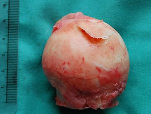
| A large flap lesion in the femur head typical of late stage Osteochondritis dissecans. In this case, the lesion was caused by avascular necrosis of the bone just under the cartilage. | |
| Pronunciation | /ˌɒsti.oʊkɒnˈdraɪtɪs ˈdɪsᵻkænz/ |
| Classification and external resources | |
| Specialty | rheumatology |
| ICD-10 | M93.2 |
| ICD-9-CM | 732.7 |
| OMIM | 165800 |
| DiseasesDB | 9320 |
| eMedicine | radio/495 sports/57 orthoped/639 |
| Patient UK | Osteochondritis dissecans |
| MeSH | D010008 |
Osteochondritis dissecans (OCD or OD) is a joint disorder in which cracks form in the articular cartilage and the underlying subchondral bone. OCD usually causes pain and swelling of the affected joint which catches and locks during movement. Physical examination typically reveals an , tenderness, and a crackling sound with joint movement.
OCD is caused by blood deprivation in the subchondral bone. This loss of blood flow causes the subchondral bone to die in a process called avascular necrosis. The bone is then reabsorbed by the body, leaving the articular cartilage it supported prone to damage. The result is fragmentation (dissection) of both cartilage and bone, and the free movement of these bone and cartilage fragments within the joint space, causing pain and further damage. OCD can be difficult to diagnose because these symptoms are found with other diseases. However, the disease can be confirmed by X-rays, computed tomography (CT) or magnetic resonance imaging (MRI) scans.
Non-surgical treatment is rarely an option as the ability for articular cartilage to heal is limited. As a result, even moderate cases require some form of surgery. When possible, non-operative forms of management such as protected reduced or non-weight bearing and immobilization are used. Surgical treatment includes arthroscopic drilling of intact lesions, securing of cartilage flap lesions with pins or screws, drilling and replacement of cartilage plugs, stem cell transplantation, and joint replacement. After surgery rehabilitation is usually a two-stage process of immobilization and physical therapy. Most rehabilitation programs combine efforts to protect the joint with muscle strengthening and range of motion. During the immobilization period, isometric exercises, such as straight leg raises, are commonly used to restore muscle loss without disturbing the cartilage of the affected joint. Once the immobilization period has ended, physical therapy involves continuous passive motion (CPM) and/or low impact activities, such as walking or swimming.
...
Wikipedia
