Odontoblasts
| Odontoblast | |
|---|---|

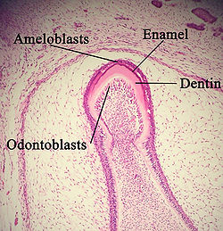
A developing tooth with odontoblasts marked.
|
|

The cervical loop area: (1) dental follicle cells, (2) dental mesenchyme, (3) Odontoblasts, (4) Dentin, (5) stellate reticulum, (6) outer enamel epithelium, (7)inner enamel epithelium, (8) ameloblasts, (9) enamel.
|
|
| Details | |
| Identifiers | |
| Latin | odontoblastus |
| Code | TE E04.0.3.3.1.0.13 |
|
Anatomical terminology
[]
|
|
In vertebrates, an odontoblast is a cell of neural crest origin that is part of the outer surface of the dental pulp, and whose biological function is dentinogenesis, which is the formation of dentin, the substance beneath the tooth enamel on the crown and the cementum on the root.
Odontoblasts first appear at sites of tooth development at 17–18 weeks in utero and remain present until death unless killed by bacterial or chemical attack, or indirectly through other means such as heat or trauma (e.g. during dental procedures). Odontoblasts were originally the outer cells of the dental papilla. Thus, dentin and pulp tissue have similar embryological backgrounds, because both are originally derived from the dental papilla of the tooth germ.
Odontoblasts are large columnar cells, whose cell bodies are arranged along the interface between dentin and pulp, from the crown to cervix to the root apex in a mature tooth. The cell is rich in endoplasmic reticulum and Golgi complex, especially during primary dentin formation, which allows it to have a high secretory capacity; it first forms the collagenous matrix to form predentin, then mineral levels to form the mature dentin. Odontoblasts form approximately 4 μm of predentin daily during tooth development.
During secretion after differentiation from the outer cells of the dental papilla, it is noted that it is polarized so its nucleus is aligned away from the newly formed dentin, with its Golgi complex and endoplasmic reticulum towards the dentin reflecting its unidirectional secretion. Thus with the formation of primary dentin, the cell moves pulpally, away from the basement membrane (future dentinoenamel junction) at the interface between the inner enamel epithelium and dental papilla, leaving behind the odontoblastic process within the pulp. The odontoblastic cell body keeps its tapered structure with cytoskeletal fibres, mainly intermediate filaments. Unlike cartilage and bone, as well as cementum, the odontoblast’s cell body does not become entrapped in the product; rather, one long, cytoplasmic attached extension remains behind in the formed dentin. The differentiation of the odontoblast is done by signaling molecules and growth factors in the cells of the inner enamel epithelium.
...
Wikipedia
