Pulp (tooth)
| Pulp (tooth) | |
|---|---|

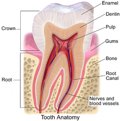
Section of a human molar
|
|
| Details | |
| Identifiers | |
| Latin | pulpa dentis |
| MeSH | A14.549.167.900.260 |
| Dorlands /Elsevier |
p_41/12679455 |
| TA | A05.1.03.051 |
| FMA | 55631 |
|
Anatomical terminology
[]
|
|
The dental pulp is the part in the center of a tooth made up of living connective tissue and cells called odontoblasts. The dental pulp is a part of the dentin–pulp complex (endodontium). The vitality of the dentin-pulp complex, both during health and after injury, depends on pulp cell activity and the signaling processes that regulate the cell’s behavior.
Each person can have a total of up to 52 pulp organs, 32 in the permanent and 20 in the primary teeth. The total volumes of all the permanent teeth organs is 0.38cc and the mean volume of a single adult human pulp is 0.02cc. Maxillary central incisor has shovel shaped coronal pulp with three short horns on the coronal roof and triangular in section. Canine has the longest pulp with elliptical cross section.
The large mass of pulp is contained within the pulp chamber of the tooth. The shape of each pulp chamber corresponds directly to the overall shape of the tooth, and thus is individualized for every tooth; the pulp tissue in the pulp chamber has two main divisions: coronal pulp and radicular pulp. Crowns of the teeth contain coronal pulp. The coronal pulp has six surfaces: the occlusal, the mesial, the distal, the buccal, the lingual and the floor. Because of continuous deposition of dentin, the pulp becomes smaller with age. This is not uniform throughout the coronal pulp but progresses faster on the floor than on the roof or side walls.
Radicular pulp is that pulp extending from the cervical region of the crown to the root apex. They are not always straight but vary in shape, size and number. The radicular portion is continuous with the periapical tissues through the apical foramen or foramina.
Apical foramen is the opening of the radicular pulp into the periapical connective tissue. The average size is 0.3 to 0.4 mm in diameter. There can be two or more foramina separated by a portion of dentin and cementum or by cementum only. If more than one foramen is present on each root, the largest one is designated as the apical foramen and the rest are considered accessory foramina. Most infections spread through the apical foramen from the pulp to periapical tissue.
...
Wikipedia
