Oculomotor
| Oculomotor nerve | |
|---|---|

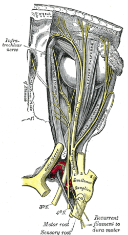
Nerves of the orbit. Seen from above.
|
|

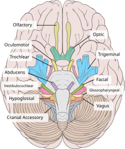
Inferior view of the human brain, with the cranial nerves labelled.
|
|
| Details | |
| From | oculomotor nucleus, Edinger-Westphal nucleus |
| To | superior branch, inferior branch |
| Innervates | Superior rectus, Inferior rectus, Medial rectus, Inferior oblique, Levator palpebrae, sphincter pupillae (parasympathetics), ciliaris muscle (parasympathetics) |
| Identifiers | |
| Latin | nervus oculomotorius |
| MeSH | A08.800.800.120.600 |
| TA | A14.2.01.007 |
| FMA | 50864 |
|
Anatomical terms of neuroanatomy
[]
|
|
The oculomotor nerve is the third cranial nerve. It enters the orbit via the superior orbital fissure and innervates muscles that enable most movements of the eye and that raise the eyelid. The nerve also contains fibers that innervate the muscles that enable pupillary constriction and accommodation (ability to focus on near objects as in reading). The oculomotor nerve is derived from the basal plate of the embryonic midbrain. Cranial nerves IV and VI also participate in control of eye movement.
The oculomotor nerve originates from the third nerve nucleus at the level of the superior colliculus in the midbrain. The third nerve nucleus is located ventral to the cerebral aqueduct, on the pre-aqueductal grey matter. The fibers from the two third nerve nuclei located laterally on either side of the cerebral aqueduct then pass through the red nucleus. From the red nucleus fibers then pass via the substantia nigra exiting through the interpeduncular fossa.
On emerging from the brainstem, the nerve is invested with a sheath of pia mater, and enclosed in a prolongation from the arachnoid. It passes between the superior cerebellar (below) and posterior cerebral arteries (above), and then pierces the dura mater anterior and lateral to the posterior clinoid process, passing between the free and attached borders of the tentorium cerebelli.
...
Wikipedia
