Myosin-light-chain phosphatase
| Myosin Light-Chain Phosphatase | |||||||||
|---|---|---|---|---|---|---|---|---|---|

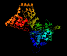
Structure of complex between PP1 and a portion of MYPT1, generated from 1s70
|
|||||||||
| Identifiers | |||||||||
| EC number | 3.1.3.53 | ||||||||
| CAS number | 86417-96-1 | ||||||||
| Databases | |||||||||
| IntEnz | IntEnz view | ||||||||
| BRENDA | BRENDA entry | ||||||||
| ExPASy | NiceZyme view | ||||||||
| KEGG | KEGG entry | ||||||||
| MetaCyc | metabolic pathway | ||||||||
| PRIAM | profile | ||||||||
| PDB structures | RCSB PDB PDBe PDBsum | ||||||||
| Gene Ontology | AmiGO / EGO | ||||||||
|
|||||||||
| Search | |
|---|---|
| PMC | articles |
| PubMed | articles |
| NCBI | proteins |
Myosin light-chain phosphatase, more commonly called myosin phosphatase (EC 3.1.3.53), is an enzyme (specifically a serine/threonine-specific protein phosphatase) that dephosphorylates the regulatory light chain of myosin II. This dephosphorylation reaction occurs in smooth muscle tissue and initiates the relaxation process of the muscle cells. Thus, myosin phosphatase undoes the muscle contraction process initiated by myosin light-chain kinase. The enzyme is composed of three subunits: the catalytic region (protein phosphatase 1, or PP1), the myosin binding subunit (MYPT1), and a third subunit (M20) of unknown function. The catalytic region uses two manganese ions as catalysts to dephosphorylate the light-chains on myosin, which causes a conformational change in the myosin and relaxes the muscle. The enzyme is highly conserved and is found in all organisms’ smooth muscle tissue. While it is known that myosin phosphatase is regulated by rho-associated protein kinases, there is current debate about whether other molecules, such as arachidonic acid and cAMP, also regulate the enzyme.
Smooth muscle tissue is mostly made of actin and myosin, two proteins that interact together to produce muscle contraction and relaxation. Myosin II, also known as conventional myosin, has two heavy chains that consist of the head and tail domains and four light chains (two per head) that bind to the heavy chains in the “neck” region. When the muscle needs to contract, calcium ions flow into the cytosol from the sarcoplasmic reticulum, where they activate calmodulin, which in turn activates myosin light-chain kinase (MLC kinase). MLC kinase phosphorylates the myosin light chain (MLC20) at the Ser-19 residue. This phosphorylation causes a conformational change in the myosin, activating crossbridge cycling and causing the muscle to contract. Because myosin undergoes a conformational change, the muscle will stay contracted even if calcium and activated MLC kinase concentrations are brought to normal levels. The conformational change must be undone to relax the muscle.
...
Wikipedia
